 Movie
Movie Controller

[English] 日本語
 Yorodumi
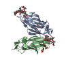
- PDB-8bxy: FimH in complex with alpha1,6 core-fucosylated oligomannose-3, cr... -
+ Open data

- Basic information
| Entry | Database: PDB / ID: 8bxy | ||||||
|---|---|---|---|---|---|---|---|
| Title | FimH in complex with alpha1,6 core-fucosylated oligomannose-3, crystallized in the trigonal space group | ||||||
 Components Components | Type 1 fimbrin D-mannose specific adhesin | ||||||
 Keywords Keywords | CELL ADHESION / adhesin / FimH / core fucose | ||||||
| Function / homology |  Function and homology information Function and homology informationpilus tip / mechanosensory behavior / Attachment of bacteria to epithelial cells / cell adhesion involved in single-species biofilm formation / pilus / cell-substrate adhesion / D-mannose binding / host cell membrane / cell adhesion Similarity search - Function | ||||||
| Biological species |  | ||||||
| Method |  X-RAY DIFFRACTION / SYNCHROTRON / MOLECULAR REPLACEMENT / Resolution: 1.45 Å X-RAY DIFFRACTION / SYNCHROTRON / MOLECULAR REPLACEMENT / Resolution: 1.45 Å | ||||||
 Authors Authors | Bridot, C. / Bouckaert, J. / Krammer, E.-M. | ||||||
| Funding support | European Union, 1items
| ||||||
 Citation Citation | Journal: J.Biol.Chem. / Year: 2023 Title: Structural insights into a cooperative switch between one and two FimH bacterial adhesins binding pauci- and high-mannose type N-glycan receptors. Authors: Krammer, E.M. / Bridot, C. / Serna, S. / Echeverria, B. / Semwal, S. / Roubinet, B. / van Noort, K. / Wilbers, R.H.P. / Bourenkov, G. / de Ruyck, J. / Landemarre, L. / Reichardt, N. / Bouckaert, J. #1: Journal: PLoS One / Year: 2008Title: Intervening with urinary tract infections using anti-adhesives based on the crystal structure of the FimH-oligomannose-3 complex. Authors: Wellens, A. / Garofalo, C. / Nguyen, H. / Van Gerven, N. / Slattegard, R. / Hernalsteens, J.P. / Wyns, L. / Oscarson, S. / De Greve, H. / Hultgren, S. / Bouckaert, J. #2: Journal: Molecules / Year: 2017Title: Sites for Dynamic Protein-Carbohydrate Interactions of O- and C-Linked Mannosides on the E. coli FimH Adhesin. Authors: Touaibia, M. / Krammer, E.M. / Shiao, T.C. / Yamakawa, N. / Wang, Q. / Glinschert, A. / Papadopoulos, A. / Mousavifar, L. / Maes, E. / Oscarson, S. / Vergoten, G. / Lensink, M.F. / Roy, R. / Bouckaert, J. #3: Journal: Molecules / Year: 2018Title: A Novel Integrated Way for Deciphering the Glycan Code for the FimH Lectin. Authors: Dumych, T. / Bridot, C. / Gouin, S.G. / Lensink, M.F. / Paryzhak, S. / Szunerits, S. / Blossey, R. / Bilyy, R. / Bouckaert, J. / Krammer, E.M. | ||||||
| History |
|
- Structure visualization
| Structure viewer | Molecule: MolmilJmol/JSmol |
|---|
- Downloads & links
-Download
| PDBx/mmCIF format | 8bxy.cif.gz | 172.9 KB | Display | PDBx/mmCIF format |
|---|---|---|---|---|
| PDB format | pdb8bxy.ent.gz | 126.3 KB | Display | PDB format |
| PDBx/mmJSON format | 8bxy.json.gz | Tree view | PDBx/mmJSON format | |
| Others |  Other downloads Other downloads |
-Validation report
| Arichive directory | https://data.pdbj.org/pub/pdb/validation_reports/bx/8bxyftp://data.pdbj.org/pub/pdb/validation_reports/bx/8bxy | HTTPS FTP |
|---|
-Related structure data
-Links
 PDBj
PDBj- Assembly
| Deposited unit | 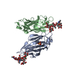
| ||||||||||||
|---|---|---|---|---|---|---|---|---|---|---|---|---|---|
| 1 | 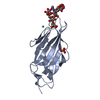
| ||||||||||||
| 2 | 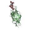
| ||||||||||||
| Unit cell |
| ||||||||||||
| Noncrystallographic symmetry (NCS) | NCS domain:
NCS domain segments: Component-ID: 1 / Ens-ID: 1 / Beg auth comp-ID: PHE / Beg label comp-ID: PHE / End auth comp-ID: THR / End label comp-ID: THR / Refine code: 1 / Auth asym-ID: A / Label asym-ID: A / Auth seq-ID: 1 - 158 / Label seq-ID: 1 - 158
NCS ensembles : (Details: Local NCS retraints between domains: 1 2) |
-Components
| #1: Protein | Mass: 16916.828 Da / Num. of mol.: 2 Source method: isolated from a genetically manipulated source Source: (gene. exp.) #2: Polysaccharide | Source method: isolated from a genetically manipulated source #3: Chemical |   Mass: 58.693 Da / Num. of mol.: 2 / Source method: obtained synthetically / Formula: Ni / Feature type: SUBJECT OF INVESTIGATION Mass: 58.693 Da / Num. of mol.: 2 / Source method: obtained synthetically / Formula: Ni / Feature type: SUBJECT OF INVESTIGATION#4: Chemical | ChemComp-SO4 / |   Mass: 96.063 Da / Num. of mol.: 1 / Source method: obtained synthetically / Formula: SO4 / Feature type: SUBJECT OF INVESTIGATION Mass: 96.063 Da / Num. of mol.: 1 / Source method: obtained synthetically / Formula: SO4 / Feature type: SUBJECT OF INVESTIGATION#5: Water | ChemComp-HOH / |  Mass: 18.015 Da / Num. of mol.: 345 / Source method: isolated from a natural source / Formula: H2O Mass: 18.015 Da / Num. of mol.: 345 / Source method: isolated from a natural source / Formula: H2OHas ligand of interest | Y | Has protein modification | Y | |
|---|
-Experimental details
-Experiment
| Experiment | Method: X-RAY DIFFRACTION / Number of used crystals: 1 |
|---|
- Sample preparation
| Crystal | Density Matthews: 2.65 Å3/Da / Density % sol: 53.66 % / Description: diamond-shaped |
|---|---|
| Crystal grow | Temperature: 291 K / Method: vapor diffusion, sitting drop / pH: 9 Details: 1.1 M Lithium sulphate; 0.1 M Tris-HCl pH 9.0; 10 mM nickel chloride |
-Data collection
| Diffraction | Mean temperature: 100 K / Serial crystal experiment: N |
|---|---|
| Diffraction source | Source: SYNCHROTRON / Site: SOLEIL  / Beamline: PROXIMA 2 / Wavelength: 0.987 Å / Beamline: PROXIMA 2 / Wavelength: 0.987 Å |
| Detector | Type: DECTRIS EIGER X 16M / Detector: PIXEL / Date: Nov 28, 2018 |
| Radiation | Protocol: SINGLE WAVELENGTH / Monochromatic (M) / Laue (L): M / Scattering type: x-ray |
| Radiation wavelength | Wavelength: 0.987 Å / Relative weight: 1 |
| Reflection | Resolution: 1.45→56.076 Å / Num. obs: 56199 / % possible obs: 95.16 % / Redundancy: 20.11 % / Biso Wilson estimate: 24.99 Å2 / CC1/2: 0.9996 / Rmerge(I) obs: 0.075 / Rpim(I) all: 0.017 / Rrim(I) all: 0.077 / Net I/σ(I): 19.325 |
| Reflection shell | Resolution: 1.45→1.556 Å / Redundancy: 16.95 % / Rmerge(I) obs: 1.837 / Mean I/σ(I) obs: 1.6 / Num. unique obs: 2811 / CC1/2: 0.615 / Rpim(I) all: 0.639 / Rrim(I) all: 1.949 / % possible all: 21.9 |
- Processing
| Software |
| |||||||||||||||||||||||||||||||||||||||||||||||||||||||||||||||||||||||||||||||||||||||||||||||||||||||||||||||||||||||||||||||||||||||||||||||||||||||||||||||||||||||||||||||||||||||||||||||||||||||||||||||||||||||||||||||||||||||
|---|---|---|---|---|---|---|---|---|---|---|---|---|---|---|---|---|---|---|---|---|---|---|---|---|---|---|---|---|---|---|---|---|---|---|---|---|---|---|---|---|---|---|---|---|---|---|---|---|---|---|---|---|---|---|---|---|---|---|---|---|---|---|---|---|---|---|---|---|---|---|---|---|---|---|---|---|---|---|---|---|---|---|---|---|---|---|---|---|---|---|---|---|---|---|---|---|---|---|---|---|---|---|---|---|---|---|---|---|---|---|---|---|---|---|---|---|---|---|---|---|---|---|---|---|---|---|---|---|---|---|---|---|---|---|---|---|---|---|---|---|---|---|---|---|---|---|---|---|---|---|---|---|---|---|---|---|---|---|---|---|---|---|---|---|---|---|---|---|---|---|---|---|---|---|---|---|---|---|---|---|---|---|---|---|---|---|---|---|---|---|---|---|---|---|---|---|---|---|---|---|---|---|---|---|---|---|---|---|---|---|---|---|---|---|---|---|---|---|---|---|---|---|---|---|---|---|---|---|---|---|---|---|
| Refinement | Method to determine structure: MOLECULAR REPLACEMENT / Resolution: 1.45→56.076 Å / Cor.coef. Fo:Fc: 0.975 / Cor.coef. Fo:Fc free: 0.959 / Cross valid method: FREE R-VALUE / ESU R: 0.076 / ESU R Free: 0.074 Details: Hydrogens have been used if present in the input file
| |||||||||||||||||||||||||||||||||||||||||||||||||||||||||||||||||||||||||||||||||||||||||||||||||||||||||||||||||||||||||||||||||||||||||||||||||||||||||||||||||||||||||||||||||||||||||||||||||||||||||||||||||||||||||||||||||||||||
| Solvent computation | Ion probe radii: 0.8 Å / Shrinkage radii: 0.8 Å / VDW probe radii: 1.2 Å / Solvent model: MASK BULK SOLVENT | |||||||||||||||||||||||||||||||||||||||||||||||||||||||||||||||||||||||||||||||||||||||||||||||||||||||||||||||||||||||||||||||||||||||||||||||||||||||||||||||||||||||||||||||||||||||||||||||||||||||||||||||||||||||||||||||||||||||
| Displacement parameters | Biso mean: 32.691 Å2
| |||||||||||||||||||||||||||||||||||||||||||||||||||||||||||||||||||||||||||||||||||||||||||||||||||||||||||||||||||||||||||||||||||||||||||||||||||||||||||||||||||||||||||||||||||||||||||||||||||||||||||||||||||||||||||||||||||||||
| Refinement step | Cycle: LAST / Resolution: 1.45→56.076 Å
| |||||||||||||||||||||||||||||||||||||||||||||||||||||||||||||||||||||||||||||||||||||||||||||||||||||||||||||||||||||||||||||||||||||||||||||||||||||||||||||||||||||||||||||||||||||||||||||||||||||||||||||||||||||||||||||||||||||||
| Refine LS restraints |
| |||||||||||||||||||||||||||||||||||||||||||||||||||||||||||||||||||||||||||||||||||||||||||||||||||||||||||||||||||||||||||||||||||||||||||||||||||||||||||||||||||||||||||||||||||||||||||||||||||||||||||||||||||||||||||||||||||||||
| Refine LS restraints NCS |
| |||||||||||||||||||||||||||||||||||||||||||||||||||||||||||||||||||||||||||||||||||||||||||||||||||||||||||||||||||||||||||||||||||||||||||||||||||||||||||||||||||||||||||||||||||||||||||||||||||||||||||||||||||||||||||||||||||||||
| LS refinement shell | Refine-ID: X-RAY DIFFRACTION / Total num. of bins used: 20
|
