 Movie
Movie Controller

[English] 日本語
 Yorodumi
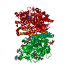
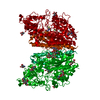
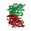
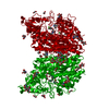
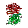
- PDB-7bfz: X-ray structure of human prostate-specific membrane antigen(PSMA)... -
+ Open data

- Basic information
| Entry | Database: PDB / ID: 7bfz | ||||||
|---|---|---|---|---|---|---|---|
| Title | X-ray structure of human prostate-specific membrane antigen(PSMA) in complex with a inhibitor Glu-490 | ||||||
 Components Components | Glutamate carboxypeptidase 2 | ||||||
 Keywords Keywords | HYDROLASE / glutamate carboxypeptidase II (GCPII) / NAALADase / prostate-specific membrane antigen (PSMA) / inhibitor | ||||||
| Function / homology |  Function and homology information Function and homology informationAc-Asp-Glu binding / tetrahydrofolyl-poly(glutamate) polymer binding / glutamate carboxypeptidase II / folic acid-containing compound metabolic process / C-terminal protein deglutamylation / Aspartate and asparagine metabolism / dipeptidase activity / carboxypeptidase activity / metallocarboxypeptidase activity / peptidase activity ...Ac-Asp-Glu binding / tetrahydrofolyl-poly(glutamate) polymer binding / glutamate carboxypeptidase II / folic acid-containing compound metabolic process / C-terminal protein deglutamylation / Aspartate and asparagine metabolism / dipeptidase activity / carboxypeptidase activity / metallocarboxypeptidase activity / peptidase activity / cell surface / proteolysis / extracellular exosome / metal ion binding / membrane / plasma membrane / cytoplasm Similarity search - Function | ||||||
| Biological species |  Homo sapiens (human) Homo sapiens (human) | ||||||
| Method |  X-RAY DIFFRACTION / SYNCHROTRON / FOURIER SYNTHESIS / Resolution: 1.73 Å X-RAY DIFFRACTION / SYNCHROTRON / FOURIER SYNTHESIS / Resolution: 1.73 Å | ||||||
 Authors Authors | Rakhimbekova, A. / Motlova, L. / Barinka, C. | ||||||
 Citation Citation | Journal: Nat Commun / Year: 2021 Title: A prostate-specific membrane antigen activated molecular rotor for real-time fluorescence imaging. Authors: Zhang, J. / Rakhimbekova, A. / Duan, X. / Yin, Q. / Foss, C.A. / Fan, Y. / Xu, Y. / Li, X. / Cai, X. / Kutil, Z. / Wang, P. / Yang, Z. / Zhang, N. / Pomper, M.G. / Wang, Y. / Barinka, C. / Yang, X. | ||||||
| History |
|
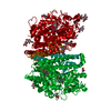
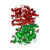
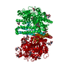
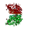
- Structure visualization
| Structure viewer | Molecule: MolmilJmol/JSmol |
|---|
- Downloads & links
-Download
| PDBx/mmCIF format | 7bfz.cif.gz | 320.7 KB | Display | PDBx/mmCIF format |
|---|---|---|---|---|
| PDB format | pdb7bfz.ent.gz | 255.9 KB | Display | PDB format |
| PDBx/mmJSON format | 7bfz.json.gz | Tree view | PDBx/mmJSON format | |
| Others |  Other downloads Other downloads |
-Validation report
| Arichive directory | https://data.pdbj.org/pub/pdb/validation_reports/bf/7bfzftp://data.pdbj.org/pub/pdb/validation_reports/bf/7bfz | HTTPS FTP |
|---|
-Related structure data
| Related structure data | 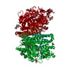 3bi1S S: Starting model for refinement |
|---|---|
| Similar structure data |


-Links
 PDBj
PDBj
- Assembly
| Deposited unit | 
| ||||||||
|---|---|---|---|---|---|---|---|---|---|
| 1 | 
| ||||||||
| Unit cell |
|
-Components
-Protein , 1 types, 1 molecules A
| #1: Protein | Mass: 79614.750 Da / Num. of mol.: 1 Source method: isolated from a genetically manipulated source Details: The missing amino acids were not possible to model into electron density map due to their flexibility. Source: (gene. exp.) Homo sapiens (human) / Gene: FOLH1, FOLH, NAALAD1, PSM, PSMA, GIG27 / Production host:  |
|---|
-Sugars , 5 types, 7 molecules 
| #2: Polysaccharide | Source method: isolated from a genetically manipulated source #3: Polysaccharide | alpha-D-mannopyranose-(1-3)-[alpha-D-mannopyranose-(1-6)]beta-D-mannopyranose-(1-4)-2-acetamido-2- ...alpha-D-mannopyranose-(1-3)-[alpha-D-mannopyranose-(1-6)]beta-D-mannopyranose-(1-4)-2-acetamido-2-deoxy-beta-D-glucopyranose-(1-4)-2-acetamido-2-deoxy-beta-D-glucopyranose | Source method: isolated from a genetically manipulated source #4: Polysaccharide | 2-acetamido-2-deoxy-beta-D-glucopyranose-(1-4)-2-acetamido-2-deoxy-beta-D-glucopyranose | Source method: isolated from a genetically manipulated source #5: Polysaccharide | beta-D-mannopyranose-(1-4)-2-acetamido-2-deoxy-beta-D-glucopyranose-(1-4)-[alpha-L-fucopyranose-(1- ...beta-D-mannopyranose-(1-4)-2-acetamido-2-deoxy-beta-D-glucopyranose-(1-4)-[alpha-L-fucopyranose-(1-6)]2-acetamido-2-deoxy-beta-D-glucopyranose | Source method: isolated from a genetically manipulated source #9: Sugar |  Type: D-saccharide, beta linking / Mass: 221.208 Da / Num. of mol.: 2 / Source method: obtained synthetically / Formula: C8H15NO6 Type: D-saccharide, beta linking / Mass: 221.208 Da / Num. of mol.: 2 / Source method: obtained synthetically / Formula: C8H15NO6 |
|---|
-Non-polymers , 8 types, 478 molecules 


| #6: Chemical |  Mass: 65.409 Da / Num. of mol.: 2 Mass: 65.409 Da / Num. of mol.: 2Source method: isolated from a genetically manipulated source Formula: Zn #7: Chemical | ChemComp-CA / |  Mass: 40.078 Da / Num. of mol.: 1 / Source method: obtained synthetically / Formula: Ca Mass: 40.078 Da / Num. of mol.: 1 / Source method: obtained synthetically / Formula: Ca#8: Chemical | ChemComp-CL / |  Mass: 35.453 Da / Num. of mol.: 1 / Source method: obtained synthetically / Formula: Cl Mass: 35.453 Da / Num. of mol.: 1 / Source method: obtained synthetically / Formula: Cl#10: Chemical |  Mass: 62.068 Da / Num. of mol.: 3 / Source method: obtained synthetically / Formula: C2H6O2 Mass: 62.068 Da / Num. of mol.: 3 / Source method: obtained synthetically / Formula: C2H6O2#11: Chemical | ChemComp-PEG / |  Mass: 106.120 Da / Num. of mol.: 1 / Source method: obtained synthetically / Formula: C4H10O3 Mass: 106.120 Da / Num. of mol.: 1 / Source method: obtained synthetically / Formula: C4H10O3#12: Chemical |  Mass: 22.990 Da / Num. of mol.: 2 / Source method: obtained synthetically / Formula: Na Mass: 22.990 Da / Num. of mol.: 2 / Source method: obtained synthetically / Formula: Na#13: Chemical | ChemComp-TKZ / ((( |  Mass: 697.755 Da / Num. of mol.: 1 / Source method: obtained synthetically / Formula: C33H39N5O10S / Feature type: SUBJECT OF INVESTIGATION Mass: 697.755 Da / Num. of mol.: 1 / Source method: obtained synthetically / Formula: C33H39N5O10S / Feature type: SUBJECT OF INVESTIGATION#14: Water | ChemComp-HOH / | Mass: 18.015 Da / Num. of mol.: 467 / Source method: isolated from a natural source / Formula: H2O |
|---|
-Details
| Has ligand of interest | Y |
|---|---|
| Has protein modification | Y |
-Experimental details
-Experiment
| Experiment | Method: X-RAY DIFFRACTION / Number of used crystals: 1 |
|---|
- Sample preparation
| Crystal | Density Matthews: 3.3 Å3/Da / Density % sol: 62.68 % |
|---|---|
| Crystal grow | Temperature: 293 K / Method: vapor diffusion, hanging drop / pH: 8 Details: 34% (v/v) pentaerythritol propoxylate PO/OH 5/4 1 % (w/v) PEG 3350 100 mM Tris-HCl, pH 8.0 |
-Data collection
| Diffraction | Mean temperature: 100 K / Serial crystal experiment: N |
|---|---|
| Diffraction source | Source: SYNCHROTRON / Site: BESSY  / Beamline: 14.2 / Wavelength: 0.9184 Å / Beamline: 14.2 / Wavelength: 0.9184 Å |
| Detector | Type: DECTRIS PILATUS3 S 2M / Detector: PIXEL / Date: Mar 1, 2020 |
| Radiation | Protocol: SINGLE WAVELENGTH / Monochromatic (M) / Laue (L): M / Scattering type: x-ray |
| Radiation wavelength | Wavelength: 0.9184 Å / Relative weight: 1 |
| Reflection | Resolution: 1.73→50 Å / Num. obs: 109170 / % possible obs: 99.6 % / Redundancy: 4.5 % / CC1/2: 0.999 / Net I/σ(I): 14.43 |
| Reflection shell | Resolution: 1.73→1.83 Å / Redundancy: 4.2 % / Mean I/σ(I) obs: 1.99 / Num. unique obs: 17360 / CC1/2: 0.869 / % possible all: 98.9 |
- Processing
| Software |
| |||||||||||||||||||||||||||||||||||||||||||||
|---|---|---|---|---|---|---|---|---|---|---|---|---|---|---|---|---|---|---|---|---|---|---|---|---|---|---|---|---|---|---|---|---|---|---|---|---|---|---|---|---|---|---|---|---|---|---|
| Refinement | Method to determine structure: FOURIER SYNTHESIS Starting model: 3BI1 Resolution: 1.73→46.96 Å / Cor.coef. Fo:Fc: 0.972 / Cor.coef. Fo:Fc free: 0.966 / SU B: 3.497 / SU ML: 0.059 / Cross valid method: THROUGHOUT / σ(F): 0 / ESU R: 0.082 / ESU R Free: 0.08 / Stereochemistry target values: MAXIMUM LIKELIHOOD / Details: U VALUES : WITH TLS ADDED
| |||||||||||||||||||||||||||||||||||||||||||||
| Solvent computation | Ion probe radii: 0.8 Å / Shrinkage radii: 0.8 Å / VDW probe radii: 1.2 Å / Solvent model: BABINET MODEL WITH MASK | |||||||||||||||||||||||||||||||||||||||||||||
| Displacement parameters | Biso max: 124.88 Å2 / Biso mean: 36.467 Å2 / Biso min: 15.82 Å2
| |||||||||||||||||||||||||||||||||||||||||||||
| Refinement step | Cycle: final / Resolution: 1.73→46.96 Å
| |||||||||||||||||||||||||||||||||||||||||||||
| Refine LS restraints |
| |||||||||||||||||||||||||||||||||||||||||||||
| LS refinement shell | Resolution: 1.73→1.774 Å / Rfactor Rfree error: 0
| |||||||||||||||||||||||||||||||||||||||||||||
| Refinement TLS params. | Method: refined / Origin x: 17.7568 Å / Origin y: 49.355 Å / Origin z: 44.7393 Å
|
