 Movie
Movie Controller

[English] 日本語
 Yorodumi
- PDB-2pvw: A high resolution structure of human glutamate carboxypeptidase I... -
+ Open data

- Basic information
| Entry | Database: PDB / ID: 2pvw | |||||||||
|---|---|---|---|---|---|---|---|---|---|---|
| Title | A high resolution structure of human glutamate carboxypeptidase II (GCPII) in complex with 2-(phosphonomethyl)pentanedioic acid (2-PMPA) | |||||||||
 Components Components | Glutamate carboxypeptidase 2 | |||||||||
 Keywords Keywords | HYDROLASE / prostate specific membrane antigen / metallopeptidase / folate hydrolase / glutamate carboxypeptidase II / Naaladase / 2-(phosphonomethyl)pentanedioic acid / 2-(PMPA) | |||||||||
| Function / homology |  Function and homology information Function and homology informationintestinal folate absorption / Ac-Asp-Glu binding / tetrahydrofolyl-poly(glutamate) polymer binding / glutamate carboxypeptidase II / folic acid-containing compound metabolic process / Aspartate and asparagine metabolism / dipeptidase activity / : / positive regulation of glutamate receptor signaling pathway / carboxypeptidase activity ...intestinal folate absorption / Ac-Asp-Glu binding / tetrahydrofolyl-poly(glutamate) polymer binding / glutamate carboxypeptidase II / folic acid-containing compound metabolic process / Aspartate and asparagine metabolism / dipeptidase activity / : / positive regulation of glutamate receptor signaling pathway / carboxypeptidase activity / metallocarboxypeptidase activity / peptidase activity / cell surface / proteolysis / extracellular exosome / membrane / metal ion binding / plasma membrane / cytoplasm Similarity search - Function | |||||||||
| Biological species |  Homo sapiens (human) Homo sapiens (human) | |||||||||
| Method |  X-RAY DIFFRACTION / SYNCHROTRON / FOURIER SYNTHESIS / Resolution: 1.71 Å X-RAY DIFFRACTION / SYNCHROTRON / FOURIER SYNTHESIS / Resolution: 1.71 Å | |||||||||
 Authors Authors | Barinka, C. / Lubkowski, J. | |||||||||
 Citation Citation | Journal: J.Med.Chem. / Year: 2007 Title: Structural insight into the pharmacophore pocket of human glutamate carboxypeptidase II. Authors: Barinka, C. / Rovenska, M. / Mlcochova, P. / Hlouchova, K. / Plechanovova, A. / Majer, P. / Tsukamoto, T. / Slusher, B.S. / Konvalinka, J. / Lubkowski, J. | |||||||||
| History |
|
- Structure visualization
| Structure viewer | Molecule: MolmilJmol/JSmol |
|---|
- Downloads & links
-Download
| PDBx/mmCIF format | 2pvw.cif.gz | 172.6 KB | Display | PDBx/mmCIF format |
|---|---|---|---|---|
| PDB format | pdb2pvw.ent.gz | 131.7 KB | Display | PDB format |
| PDBx/mmJSON format | 2pvw.json.gz | Tree view | PDBx/mmJSON format | |
| Others |  Other downloads Other downloads |
-Validation report
| Arichive directory | https://data.pdbj.org/pub/pdb/validation_reports/pv/2pvwftp://data.pdbj.org/pub/pdb/validation_reports/pv/2pvw | HTTPS FTP |
|---|
-Related structure data
| Related structure data |  2or4C  2pvvC  2ootS S: Starting model for refinement C: citing same article ( |
|---|---|
| Similar structure data |


-Links
 PDBj
PDBj
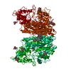
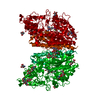
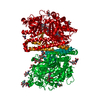
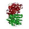
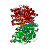
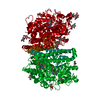
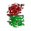
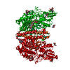
- Assembly
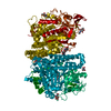
| Deposited unit | 
| ||||||||
|---|---|---|---|---|---|---|---|---|---|
| 1 | 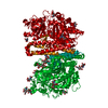
| ||||||||
| Unit cell |
| ||||||||
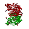
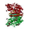
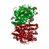
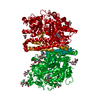
| Details | Biological assembly is a dimer. There is one molecule of GCPII in an asymmetric unit. The biological dimer is formed using "-x; 1-y; z" symmetry operator. |
-Components
-Protein , 1 types, 1 molecules A
| #1: Protein | Mass: 79859.031 Da / Num. of mol.: 1 / Fragment: extracellular domain Source method: isolated from a genetically manipulated source Source: (gene. exp.) Homo sapiens (human) / Gene: FOLH1, FOLH, NAALAD1, PSM, PSMA / Plasmid: pMT/BiP/V5-His A / Production host:  |
|---|
-Sugars , 3 types, 7 molecules 
| #2: Polysaccharide | Source method: isolated from a genetically manipulated source #3: Polysaccharide | alpha-D-mannopyranose-(1-3)-beta-D-mannopyranose-(1-4)-2-acetamido-2-deoxy-beta-D-glucopyranose-(1- ...alpha-D-mannopyranose-(1-3)-beta-D-mannopyranose-(1-4)-2-acetamido-2-deoxy-beta-D-glucopyranose-(1-4)-2-acetamido-2-deoxy-beta-D-glucopyranose | Source method: isolated from a genetically manipulated source #4: Sugar |  Type: D-saccharide, beta linking / Mass: 221.208 Da / Num. of mol.: 3 Type: D-saccharide, beta linking / Mass: 221.208 Da / Num. of mol.: 3Source method: isolated from a genetically manipulated source Formula: C8H15NO6 |
|---|
-Non-polymers , 5 types, 426 molecules 


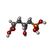

| #5: Chemical |  Mass: 65.409 Da / Num. of mol.: 2 / Source method: obtained synthetically / Formula: Zn Mass: 65.409 Da / Num. of mol.: 2 / Source method: obtained synthetically / Formula: Zn#6: Chemical | ChemComp-CA / |  Mass: 40.078 Da / Num. of mol.: 1 / Source method: obtained synthetically / Formula: Ca Mass: 40.078 Da / Num. of mol.: 1 / Source method: obtained synthetically / Formula: Ca#7: Chemical | ChemComp-CL / |  Mass: 35.453 Da / Num. of mol.: 1 / Source method: obtained synthetically / Formula: Cl Mass: 35.453 Da / Num. of mol.: 1 / Source method: obtained synthetically / Formula: Cl#8: Chemical | ChemComp-G88 / ( |  Mass: 226.121 Da / Num. of mol.: 1 / Source method: obtained synthetically / Formula: C6H11O7P Mass: 226.121 Da / Num. of mol.: 1 / Source method: obtained synthetically / Formula: C6H11O7P#9: Water | ChemComp-HOH / | Mass: 18.015 Da / Num. of mol.: 421 / Source method: isolated from a natural source / Formula: H2O |
|---|
-Details
| Has protein modification | Y |
|---|
-Experimental details
-Experiment
| Experiment | Method: X-RAY DIFFRACTION / Number of used crystals: 1 |
|---|
- Sample preparation
| Crystal | Density Matthews: 3.32 Å3/Da / Density % sol: 62.99 % |
|---|---|
| Crystal grow | Temperature: 293 K / Method: vapor diffusion, hanging drop / pH: 8 Details: 33% (v/v) pentaerythritol propoxylate PO/OH 5/4, 1 3% (w/v) PEG 3350, 100 mM Tris-HCl, pH 8.0, VAPOR DIFFUSION, HANGING DROP, temperature 293K |
-Data collection
| Diffraction | Mean temperature: 100 K |
|---|---|
| Diffraction source | Source: SYNCHROTRON / Site: APS  / Beamline: 22-ID / Wavelength: 1 Å / Beamline: 22-ID / Wavelength: 1 Å |
| Detector | Type: MARMOSAIC 300 mm CCD / Detector: CCD / Date: Jul 24, 2006 |
| Radiation | Protocol: SINGLE WAVELENGTH / Monochromatic (M) / Laue (L): M / Scattering type: x-ray |
| Radiation wavelength | Wavelength: 1 Å / Relative weight: 1 |
| Reflection | Resolution: 1.71→30 Å / Num. all: 111281 / Num. obs: 111281 / % possible obs: 98.6 % / Observed criterion σ(F): -3 / Observed criterion σ(I): -3 / Redundancy: 9.8 % / Rmerge(I) obs: 0.126 / Net I/σ(I): 15.1 |
| Reflection shell | Resolution: 1.72→1.78 Å / Redundancy: 6 % / Rmerge(I) obs: 0.495 / Mean I/σ(I) obs: 3.5 / Num. unique all: 10238 / % possible all: 91.7 |
- Processing
| Software |
| ||||||||||||||||||||||||||||||||||||||||||||||||||||||||||||||||||||||||||||||||||||||||||||||||||||||||||||||||||||||||||||||||||||||||||||||||||||||||||||||||||||||||||
|---|---|---|---|---|---|---|---|---|---|---|---|---|---|---|---|---|---|---|---|---|---|---|---|---|---|---|---|---|---|---|---|---|---|---|---|---|---|---|---|---|---|---|---|---|---|---|---|---|---|---|---|---|---|---|---|---|---|---|---|---|---|---|---|---|---|---|---|---|---|---|---|---|---|---|---|---|---|---|---|---|---|---|---|---|---|---|---|---|---|---|---|---|---|---|---|---|---|---|---|---|---|---|---|---|---|---|---|---|---|---|---|---|---|---|---|---|---|---|---|---|---|---|---|---|---|---|---|---|---|---|---|---|---|---|---|---|---|---|---|---|---|---|---|---|---|---|---|---|---|---|---|---|---|---|---|---|---|---|---|---|---|---|---|---|---|---|---|---|---|---|---|
| Refinement | Method to determine structure: FOURIER SYNTHESIS Starting model: 2OOT Resolution: 1.71→15 Å / Cor.coef. Fo:Fc: 0.968 / Cor.coef. Fo:Fc free: 0.959 / SU B: 1.898 / SU ML: 0.062 / Isotropic thermal model: mixed / Cross valid method: THROUGHOUT / ESU R: 0.089 / ESU R Free: 0.088 / Stereochemistry target values: MAXIMUM LIKELIHOOD / Details: HYDROGENS HAVE BEEN ADDED IN THE RIDING POSITIONS
| ||||||||||||||||||||||||||||||||||||||||||||||||||||||||||||||||||||||||||||||||||||||||||||||||||||||||||||||||||||||||||||||||||||||||||||||||||||||||||||||||||||||||||
| Solvent computation | Ion probe radii: 0.8 Å / Shrinkage radii: 0.8 Å / VDW probe radii: 1.2 Å / Solvent model: MASK | ||||||||||||||||||||||||||||||||||||||||||||||||||||||||||||||||||||||||||||||||||||||||||||||||||||||||||||||||||||||||||||||||||||||||||||||||||||||||||||||||||||||||||
| Displacement parameters | Biso mean: 34.647 Å2
| ||||||||||||||||||||||||||||||||||||||||||||||||||||||||||||||||||||||||||||||||||||||||||||||||||||||||||||||||||||||||||||||||||||||||||||||||||||||||||||||||||||||||||
| Refinement step | Cycle: LAST / Resolution: 1.71→15 Å
| ||||||||||||||||||||||||||||||||||||||||||||||||||||||||||||||||||||||||||||||||||||||||||||||||||||||||||||||||||||||||||||||||||||||||||||||||||||||||||||||||||||||||||
| Refine LS restraints |
| ||||||||||||||||||||||||||||||||||||||||||||||||||||||||||||||||||||||||||||||||||||||||||||||||||||||||||||||||||||||||||||||||||||||||||||||||||||||||||||||||||||||||||
| LS refinement shell | Resolution: 1.713→1.757 Å / Total num. of bins used: 20
|
