 Movie
Movie Controller

[English] 日本語
 Yorodumi
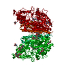
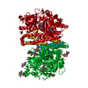
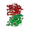
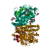
- PDB-2jbk: membrane-bound glutamate carboxypeptidase II (GCPII) in complex w... -
+ Open data

- Basic information
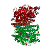
| Entry | Database: PDB / ID: 2jbk | |||||||||
|---|---|---|---|---|---|---|---|---|---|---|
| Title | membrane-bound glutamate carboxypeptidase II (GCPII) in complex with quisqualic acid (quisqualate, alpha-amino-3,5-dioxo-1,2,4- oxadiazolidine-2-propanoic acid) | |||||||||
 Components Components | GLUTAMATE CARBOXYPEPTIDASE 2 | |||||||||
 Keywords Keywords | HYDROLASE / MULTIFUNCTIONAL ENZYME / NEURODEGENERATIVE DISEASE / GLYCOPROTEIN / METAL-BINDING / SIGNAL-ANCHOR / NAALADASE / DIPEPTIDASE / POLYMORPHISM / ZINC / PSMA / ANTIGEN / MEMBRANE / PROTEASE / PEPTIDASE / TRANSMEMBRANE / METAL- BINDING / METALLOPROTEASE / PROSTATE CANCER / CARBOXYPEPTIDASE | |||||||||
| Function / homology |  Function and homology information Function and homology informationintestinal folate absorption / Ac-Asp-Glu binding / tetrahydrofolyl-poly(glutamate) polymer binding / glutamate carboxypeptidase II / folic acid-containing compound metabolic process / Aspartate and asparagine metabolism / dipeptidase activity / : / positive regulation of glutamate receptor signaling pathway / carboxypeptidase activity ...intestinal folate absorption / Ac-Asp-Glu binding / tetrahydrofolyl-poly(glutamate) polymer binding / glutamate carboxypeptidase II / folic acid-containing compound metabolic process / Aspartate and asparagine metabolism / dipeptidase activity / : / positive regulation of glutamate receptor signaling pathway / carboxypeptidase activity / metallocarboxypeptidase activity / peptidase activity / cell surface / proteolysis / extracellular exosome / membrane / metal ion binding / plasma membrane / cytoplasm Similarity search - Function | |||||||||
| Biological species |  HOMO SAPIENS (human) HOMO SAPIENS (human) | |||||||||
| Method |  X-RAY DIFFRACTION / SYNCHROTRON / MOLECULAR REPLACEMENT / Resolution: 2.99 Å X-RAY DIFFRACTION / SYNCHROTRON / MOLECULAR REPLACEMENT / Resolution: 2.99 Å | |||||||||
 Authors Authors | Mesters, J.R. / Henning, K. / Hilgenfeld, R. | |||||||||
 Citation Citation | Journal: Acta Crystallogr.,Sect.D / Year: 2007 Title: Human Glutamate Carboxypeptidase II Inhibition: Structures of Gcpii in Complex with Two Potent Inhibitors, Quisqualate and 2-Pmpa. Authors: Mesters, J.R. / Henning, K. / Hilgenfeld, R. #1: Journal: Embo J. / Year: 2006Title: Structure of Glutamate Carboxypeptidase II, a Drug Target in Neuronal Damage and Prostate Cancer Authors: Mesters, J.R. / Barinka, C. / Li, W. / Tsukamoto, T. / Majer, P. / Slusher, B.S. / Konvalinka, J. / Hilgenfeld, R. | |||||||||
| History |
|
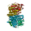
- Structure visualization
| Structure viewer | Molecule: MolmilJmol/JSmol |
|---|
- Downloads & links
-Download
| PDBx/mmCIF format | 2jbk.cif.gz | 140.4 KB | Display | PDBx/mmCIF format |
|---|---|---|---|---|
| PDB format | pdb2jbk.ent.gz | 103.1 KB | Display | PDB format |
| PDBx/mmJSON format | 2jbk.json.gz | Tree view | PDBx/mmJSON format | |
| Others |  Other downloads Other downloads |
-Validation report
| Arichive directory | https://data.pdbj.org/pub/pdb/validation_reports/jb/2jbkftp://data.pdbj.org/pub/pdb/validation_reports/jb/2jbk | HTTPS FTP |
|---|
-Related structure data
| Related structure data |  2jbjC  2c6cS S: Starting model for refinement C: citing same article ( |
|---|---|
| Similar structure data |


-Links
 PDBj
PDBj
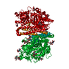
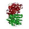
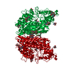
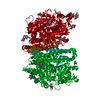
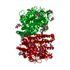
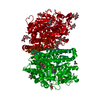
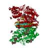
- Assembly
| Deposited unit | 
| ||||||||
|---|---|---|---|---|---|---|---|---|---|
| 1 | 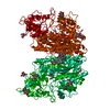
| ||||||||
| Unit cell |
|
-Components
-Protein , 1 types, 1 molecules A
| #1: Protein | Mass: 79614.750 Da / Num. of mol.: 1 / Fragment: EXTRACELLULAR PART, RESIDUES 44-750 Source method: isolated from a genetically manipulated source Source: (gene. exp.) HOMO SAPIENS (human) / Organ: BRAIN / Cell line (production host): SCHNEIDER'S CELLS / Production host:  |
|---|
-Sugars , 3 types, 7 molecules 
| #2: Polysaccharide | Source method: isolated from a genetically manipulated source #3: Polysaccharide | alpha-D-mannopyranose-(1-3)-beta-D-mannopyranose-(1-4)-2-acetamido-2-deoxy-beta-D-glucopyranose-(1- ...alpha-D-mannopyranose-(1-3)-beta-D-mannopyranose-(1-4)-2-acetamido-2-deoxy-beta-D-glucopyranose-(1-4)-2-acetamido-2-deoxy-beta-D-glucopyranose | Source method: isolated from a genetically manipulated source #7: Sugar |  Type: D-saccharide, beta linking / Mass: 221.208 Da / Num. of mol.: 3 Type: D-saccharide, beta linking / Mass: 221.208 Da / Num. of mol.: 3Source method: isolated from a genetically manipulated source Formula: C8H15NO6 |
|---|
-Non-polymers , 5 types, 26 molecules 


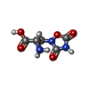

| #4: Chemical |  Mass: 65.409 Da / Num. of mol.: 2 / Source method: obtained synthetically / Formula: Zn Mass: 65.409 Da / Num. of mol.: 2 / Source method: obtained synthetically / Formula: Zn#5: Chemical | ChemComp-CA / |  Mass: 40.078 Da / Num. of mol.: 1 / Source method: obtained synthetically / Formula: Ca Mass: 40.078 Da / Num. of mol.: 1 / Source method: obtained synthetically / Formula: Ca#6: Chemical | ChemComp-CL / |  Mass: 35.453 Da / Num. of mol.: 1 / Source method: obtained synthetically / Formula: Cl Mass: 35.453 Da / Num. of mol.: 1 / Source method: obtained synthetically / Formula: Cl#8: Chemical | ChemComp-QUS / ( |  Mass: 189.126 Da / Num. of mol.: 1 / Source method: obtained synthetically / Formula: C5H7N3O5 Mass: 189.126 Da / Num. of mol.: 1 / Source method: obtained synthetically / Formula: C5H7N3O5#9: Water | ChemComp-HOH / | Mass: 18.015 Da / Num. of mol.: 21 / Source method: isolated from a natural source / Formula: H2O |
|---|
-Details
| Has protein modification | Y |
|---|---|
| Sequence details | AMINO ACIDS 44-750 |
-Experimental details
-Experiment
| Experiment | Method: X-RAY DIFFRACTION / Number of used crystals: 1 |
|---|
- Sample preparation
| Crystal | Density Matthews: 3.48 Å3/Da / Density % sol: 64.42 % / Description: NONE |
|---|---|
| Crystal grow | pH: 7 Details: 20 MM HEPES PH 7.25, 200 MM NACL, 5% (W/V) PEG 400 AND 15% (W/V) PEG 1500. |
-Data collection
| Diffraction | Mean temperature: 260 K |
|---|---|
| Diffraction source | Source: SYNCHROTRON / Site: EMBL/DESY, HAMBURG  / Beamline: X11 / Wavelength: 0.8115 / Beamline: X11 / Wavelength: 0.8115 |
| Detector | Type: MARRESEARCH / Detector: CCD / Date: Jan 27, 2004 |
| Radiation | Protocol: SINGLE WAVELENGTH / Monochromatic (M) / Laue (L): M / Scattering type: x-ray |
| Radiation wavelength | Wavelength: 0.8115 Å / Relative weight: 1 |
| Reflection | Resolution: 2.99→50 Å / Num. obs: 20662 / % possible obs: 93.5 % / Observed criterion σ(I): 0 / Redundancy: 5 % / Rmerge(I) obs: 0.15 |
- Processing
| Software |
| ||||||||||||||||||||||||||||||||||||||||||||||||||||||||||||||||||||||||||||||||||||||||||||||||||||||||||||||||||||||||||||||||||||||||||||||||||||||||||||||||||||||||||||||||||||||
|---|---|---|---|---|---|---|---|---|---|---|---|---|---|---|---|---|---|---|---|---|---|---|---|---|---|---|---|---|---|---|---|---|---|---|---|---|---|---|---|---|---|---|---|---|---|---|---|---|---|---|---|---|---|---|---|---|---|---|---|---|---|---|---|---|---|---|---|---|---|---|---|---|---|---|---|---|---|---|---|---|---|---|---|---|---|---|---|---|---|---|---|---|---|---|---|---|---|---|---|---|---|---|---|---|---|---|---|---|---|---|---|---|---|---|---|---|---|---|---|---|---|---|---|---|---|---|---|---|---|---|---|---|---|---|---|---|---|---|---|---|---|---|---|---|---|---|---|---|---|---|---|---|---|---|---|---|---|---|---|---|---|---|---|---|---|---|---|---|---|---|---|---|---|---|---|---|---|---|---|---|---|---|---|
| Refinement | Method to determine structure: MOLECULAR REPLACEMENT Starting model: PDB ENTRY 2C6C Resolution: 2.99→100 Å / Cor.coef. Fo:Fc: 0.912 / SU B: 11.469 / SU ML: 0.211 / Cross valid method: THROUGHOUT / Stereochemistry target values: MAXIMUM LIKELIHOOD / Details: HYDROGENS HAVE BEEN ADDED IN THE RIDING POSITIONS.
| ||||||||||||||||||||||||||||||||||||||||||||||||||||||||||||||||||||||||||||||||||||||||||||||||||||||||||||||||||||||||||||||||||||||||||||||||||||||||||||||||||||||||||||||||||||||
| Solvent computation | Ion probe radii: 0.8 Å / Shrinkage radii: 0.8 Å / VDW probe radii: 1.2 Å / Solvent model: MASK | ||||||||||||||||||||||||||||||||||||||||||||||||||||||||||||||||||||||||||||||||||||||||||||||||||||||||||||||||||||||||||||||||||||||||||||||||||||||||||||||||||||||||||||||||||||||
| Displacement parameters | Biso mean: 34.08 Å2
| ||||||||||||||||||||||||||||||||||||||||||||||||||||||||||||||||||||||||||||||||||||||||||||||||||||||||||||||||||||||||||||||||||||||||||||||||||||||||||||||||||||||||||||||||||||||
| Refinement step | Cycle: LAST / Resolution: 2.99→100 Å
| ||||||||||||||||||||||||||||||||||||||||||||||||||||||||||||||||||||||||||||||||||||||||||||||||||||||||||||||||||||||||||||||||||||||||||||||||||||||||||||||||||||||||||||||||||||||
| Refine LS restraints |
|