 Movie
Movie Controller

[English] 日本語
 Yorodumi
- PDB-7bcb: Crystal structure of the HTH DNA binding protein ArdK from R388 p... -
+ Open data

- Basic information
| Entry | Database: PDB / ID: 7bcb | |||||||||
|---|---|---|---|---|---|---|---|---|---|---|
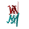
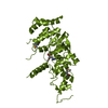
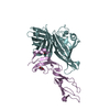
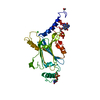
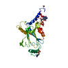
| Title | Crystal structure of the HTH DNA binding protein ArdK from R388 plasmid bound to IR3 DNA | |||||||||
 Components Components |
| |||||||||
 Keywords Keywords | DNA BINDING PROTEIN / Helix Turn Helix / Direct repeat / Plasmid conjugation | |||||||||
| Function / homology |  Function and homology information Function and homology informationArc Repressor Mutant, subunit A - #2690 / TrfB transcriptional repressor protein / TrfB plasmid transcriptional repressor / : / RNA polymerase sigma factor, region 3/4-like / Arc Repressor Mutant, subunit A / Orthogonal Bundle / Mainly Alpha Similarity search - Domain/homology | |||||||||
| Biological species |  | |||||||||
| Method |  X-RAY DIFFRACTION / SYNCHROTRON / MOLECULAR REPLACEMENT / Resolution: 2.8 Å X-RAY DIFFRACTION / SYNCHROTRON / MOLECULAR REPLACEMENT / Resolution: 2.8 Å | |||||||||
 Authors Authors | Fernandez-Lopez, R. / Boer, D.R. / Moncalian, G. | |||||||||
| Funding support |  Spain, 2items Spain, 2items
| |||||||||
 Citation Citation | Journal: Nucleic Acids Res. / Year: 2022 Title: Structural basis of direct and inverted DNA sequence repeat recognition by helix-turn-helix transcription factors. Authors: Fernandez-Lopez, R. / Ruiz, R. / Del Campo, I. / Gonzalez-Montes, L. / Boer, D.R. / de la Cruz, F. / Moncalian, G. | |||||||||
| History |
|
- Structure visualization
| Structure viewer | Molecule: MolmilJmol/JSmol |
|---|
- Downloads & links
-Download
| PDBx/mmCIF format | 7bcb.cif.gz | 86.7 KB | Display | PDBx/mmCIF format |
|---|---|---|---|---|
| PDB format | pdb7bcb.ent.gz | 49.8 KB | Display | PDB format |
| PDBx/mmJSON format | 7bcb.json.gz | Tree view | PDBx/mmJSON format | |
| Others |  Other downloads Other downloads |
-Validation report
| Summary document | 7bcb_validation.pdf.gz | 424.4 KB | Display | wwPDB validaton report |
|---|---|---|---|---|
| Full document | 7bcb_full_validation.pdf.gz | 430.1 KB | Display | |
| Data in XML | 7bcb_validation.xml.gz | 6.7 KB | Display | |
| Data in CIF | 7bcb_validation.cif.gz | 9.2 KB | Display | |
| Arichive directory | https://data.pdbj.org/pub/pdb/validation_reports/bc/7bcbftp://data.pdbj.org/pub/pdb/validation_reports/bc/7bcb | HTTPS FTP |
-Related structure data
| Related structure data | 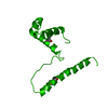 7bbqC 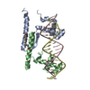 7bcaSC S: Starting model for refinement C: citing same article ( |
|---|---|
| Similar structure data |


-Links
 PDBj
PDBj


- Assembly
| Deposited unit | 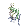
| ||||||||||||
|---|---|---|---|---|---|---|---|---|---|---|---|---|---|
| 1 |
| ||||||||||||
| Unit cell |
|
-Components
| #1: Protein | Mass: 12518.110 Da / Num. of mol.: 2 Source method: isolated from a genetically manipulated source Details: R388 plasmid / Source: (gene. exp.) #2: DNA chain | | Mass: 5522.629 Da / Num. of mol.: 1 / Source method: obtained synthetically / Source: (synth.) #3: DNA chain | | Mass: 5504.601 Da / Num. of mol.: 1 / Source method: obtained synthetically / Source: (synth.) |
|---|
-Experimental details
-Experiment
| Experiment | Method: X-RAY DIFFRACTION / Number of used crystals: 1 |
|---|
- Sample preparation
| Crystal | Density Matthews: 2.97 Å3/Da / Density % sol: 58.56 % |
|---|---|
| Crystal grow | Temperature: 295 K / Method: vapor diffusion, sitting drop / pH: 7.5 / Details: 20% 10,000 polyethylene glycol, 0.1 M HEPES pH 7.5 |
-Data collection
| Diffraction | Mean temperature: 100 K / Serial crystal experiment: N |
|---|---|
| Diffraction source | Source: SYNCHROTRON / Site: ALBA / Beamline: XALOC / Wavelength: 0.9792 Å |
| Detector | Type: DECTRIS PILATUS 6M / Detector: PIXEL / Date: Dec 3, 2016 |
| Radiation | Protocol: SINGLE WAVELENGTH / Monochromatic (M) / Laue (L): M / Scattering type: x-ray |
| Radiation wavelength | Wavelength: 0.9792 Å / Relative weight: 1 |
| Reflection | Resolution: 2.6→77.22 Å / Num. obs: 12277 / % possible obs: 98.3 % / Redundancy: 8.1 % / Biso Wilson estimate: 61.38 Å2 / CC1/2: 0.995 / Rmerge(I) obs: 0.084 / Rrim(I) all: 0.102 / Net I/σ(I): 4.6 |
| Reflection shell | Resolution: 2.6→2.74 Å / Rmerge(I) obs: 0.362 / Mean I/σ(I) obs: 4.7 / Num. unique obs: 1744 / CC1/2: 0.94 / Rrim(I) all: 0.446 |
- Processing
| Software |
| ||||||||||||||||||||||||||||
|---|---|---|---|---|---|---|---|---|---|---|---|---|---|---|---|---|---|---|---|---|---|---|---|---|---|---|---|---|---|
| Refinement | Method to determine structure: MOLECULAR REPLACEMENT Starting model: 7BCA Resolution: 2.8→45.89 Å / SU ML: 0.504 / Cross valid method: FREE R-VALUE / σ(F): 1.34 / Phase error: 38.7723 Stereochemistry target values: GeoStd + Monomer Library + CDL v1.2
| ||||||||||||||||||||||||||||
| Solvent computation | Shrinkage radii: 0.9 Å / VDW probe radii: 1.1 Å / Solvent model: FLAT BULK SOLVENT MODEL | ||||||||||||||||||||||||||||
| Displacement parameters | Biso mean: 66.08 Å2 | ||||||||||||||||||||||||||||
| Refinement step | Cycle: LAST / Resolution: 2.8→45.89 Å
| ||||||||||||||||||||||||||||
| Refine LS restraints |
| ||||||||||||||||||||||||||||
| LS refinement shell |
|
