 Movie
Movie Controller

[English] 日本語
 Yorodumi
- PDB-2oh7: The Crystal Structure of Cypovirus Polyhedra containing the Human... -
+ Open data

- Basic information
| Entry | Database: PDB / ID: 2oh7 | ||||||
|---|---|---|---|---|---|---|---|
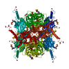
| Title | The Crystal Structure of Cypovirus Polyhedra containing the Human ZIP-kinase | ||||||
 Components Components | Polyhedrin | ||||||
 Keywords Keywords | Structural Protein / RNA binding protein / beta sandwich / intracellular crystal / nucleotide binding | ||||||
| Function / homology |  Function and homology information Function and homology informationGTP binding / ATP binding / metal ion binding / identical protein binding Similarity search - Function | ||||||
| Biological species |  | ||||||
| Method |  X-RAY DIFFRACTION / SYNCHROTRON / MOLECULAR REPLACEMENT / Resolution: 2.45 Å X-RAY DIFFRACTION / SYNCHROTRON / MOLECULAR REPLACEMENT / Resolution: 2.45 Å | ||||||
 Authors Authors | Coulibaly, F. / Chiu, E. / Ikeda, K. / Gutmann, S. / Haebel, P.W. / Schulze-Briese, C. / Mori, H. / Metcalf, P. | ||||||
 Citation Citation | Journal: Nature / Year: 2007 Title: The molecular organization of cypovirus polyhedra. Authors: Coulibaly, F. / Chiu, E. / Ikeda, K. / Gutmann, S. / Haebel, P.W. / Schulze-Briese, C. / Mori, H. / Metcalf, P. | ||||||
| History |
|
- Structure visualization
| Structure viewer | Molecule: MolmilJmol/JSmol |
|---|
- Downloads & links
-Download
| PDBx/mmCIF format | 2oh7.cif.gz | 71.1 KB | Display | PDBx/mmCIF format |
|---|---|---|---|---|
| PDB format | pdb2oh7.ent.gz | 51.2 KB | Display | PDB format |
| PDBx/mmJSON format | 2oh7.json.gz | Tree view | PDBx/mmJSON format | |
| Others |  Other downloads Other downloads |
-Validation report
| Arichive directory | https://data.pdbj.org/pub/pdb/validation_reports/oh/2oh7ftp://data.pdbj.org/pub/pdb/validation_reports/oh/2oh7 | HTTPS FTP |
|---|
-Related structure data
| Related structure data | 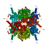 2oh5C 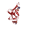 2oh6SC C: citing same article ( S: Starting model for refinement |
|---|---|
| Similar structure data |


-Links
 PDBj
PDBj
- Assembly
| Deposited unit | 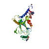
| |||||||||
|---|---|---|---|---|---|---|---|---|---|---|
| 1 | x 12
| |||||||||
| Unit cell |
| |||||||||
| Components on special symmetry positions |
| |||||||||
| Details | The whole micron-sized crystals, called polyhedra, are the biological units. Polyhedra crystallize in the cytoplasm of CPV-infected cells and protect the virus particles. |
-Components
-Protein , 1 types, 1 molecules A
| #1: Protein | Mass: 28360.355 Da / Num. of mol.: 1 / Mutation: N29S Source method: isolated from a genetically manipulated source Source: (gene. exp.)  Spodoptera frugiperda (fall armyworm) / Strain (production host): Sf21 (IPLB-SF21AE) / References: UniProt: O10693 Spodoptera frugiperda (fall armyworm) / Strain (production host): Sf21 (IPLB-SF21AE) / References: UniProt: O10693 |
|---|
-Non-polymers , 6 types, 143 molecules 


| #2: Chemical | ChemComp-CL /  Mass: 35.453 Da / Num. of mol.: 1 / Source method: obtained synthetically / Formula: Cl Mass: 35.453 Da / Num. of mol.: 1 / Source method: obtained synthetically / Formula: Cl | ||||||||
|---|---|---|---|---|---|---|---|---|---|
| #3: Chemical |  Mass: 24.305 Da / Num. of mol.: 2 / Source method: obtained synthetically / Formula: Mg Mass: 24.305 Da / Num. of mol.: 2 / Source method: obtained synthetically / Formula: Mg#4: Chemical | ChemComp-GTP / |  Mass: 523.180 Da / Num. of mol.: 1 / Source method: obtained synthetically / Formula: C10H16N5O14P3 / Comment: GTP, energy-carrying molecule*YM Mass: 523.180 Da / Num. of mol.: 1 / Source method: obtained synthetically / Formula: C10H16N5O14P3 / Comment: GTP, energy-carrying molecule*YM#5: Chemical | ChemComp-ATP / |  Mass: 507.181 Da / Num. of mol.: 1 / Source method: obtained synthetically / Formula: C10H16N5O13P3 / Comment: ATP, energy-carrying molecule*YM Mass: 507.181 Da / Num. of mol.: 1 / Source method: obtained synthetically / Formula: C10H16N5O13P3 / Comment: ATP, energy-carrying molecule*YM#6: Chemical | ChemComp-CTP / |  Mass: 483.156 Da / Num. of mol.: 1 / Source method: obtained synthetically / Formula: C9H16N3O14P3 Mass: 483.156 Da / Num. of mol.: 1 / Source method: obtained synthetically / Formula: C9H16N3O14P3#7: Water | ChemComp-HOH / | Mass: 18.015 Da / Num. of mol.: 137 / Source method: isolated from a natural source / Formula: H2O |
-Details
| Has protein modification | Y |
|---|
-Experimental details
-Experiment
| Experiment | Method: X-RAY DIFFRACTION / Number of used crystals: 5 |
|---|
- Sample preparation
| Crystal grow | Temperature: 300 K Details: The crystals used to determine this structure were directly purified from cells, in vivo crystallization in the cytoplasm of the cell, temperature 300K |
|---|
-Data collection
| Diffraction | Mean temperature: 120 K |
|---|---|
| Diffraction source | Source: SYNCHROTRON / Site: SLS  / Beamline: X06SA / Wavelength: 0.9983 Å / Beamline: X06SA / Wavelength: 0.9983 Å |
| Detector | Type: MAR CCD 165 mm / Detector: CCD / Date: Aug 5, 2006 / Details: MD2 diffractometer |
| Radiation | Monochromator: sagitally horizontal focussing Si(111); meridionally vertical focussing Rh-coated mirror Protocol: SINGLE WAVELENGTH / Monochromatic (M) / Laue (L): M / Scattering type: x-ray |
| Radiation wavelength | Wavelength: 0.9983 Å / Relative weight: 1 |
| Reflection | Resolution: 2.45→20 Å / Num. all: 6778 / Num. obs: 6770 / % possible obs: 99.9 % / Observed criterion σ(I): -3 / Redundancy: 8.2 % / Biso Wilson estimate: 23.4 Å2 / Rmerge(I) obs: 0.128 / Χ2: 1.025 / Net I/σ(I): 8.2 |
| Reflection shell | Resolution: 2.45→2.54 Å / Redundancy: 3.5 % / Rmerge(I) obs: 0.348 / Mean I/σ(I) obs: 3.7 / Num. unique all: 656 / Χ2: 1.08 / % possible all: 99.4 |
- Processing
| Software |
| |||||||||||||||||||||||||||||||||||||||||||||||||||||||||||||||||||||||||||||||||||||||||||||||||||||||||||||||||||||||||||||
|---|---|---|---|---|---|---|---|---|---|---|---|---|---|---|---|---|---|---|---|---|---|---|---|---|---|---|---|---|---|---|---|---|---|---|---|---|---|---|---|---|---|---|---|---|---|---|---|---|---|---|---|---|---|---|---|---|---|---|---|---|---|---|---|---|---|---|---|---|---|---|---|---|---|---|---|---|---|---|---|---|---|---|---|---|---|---|---|---|---|---|---|---|---|---|---|---|---|---|---|---|---|---|---|---|---|---|---|---|---|---|---|---|---|---|---|---|---|---|---|---|---|---|---|---|---|---|
| Refinement | Method to determine structure: MOLECULAR REPLACEMENT Starting model: PDB entry 2OH6 Resolution: 2.45→18.8 Å / Cor.coef. Fo:Fc: 0.974 / Cor.coef. Fo:Fc free: 0.908 / SU B: 8.773 / SU ML: 0.2 / Isotropic thermal model: Isotropic / Cross valid method: THROUGHOUT / σ(F): 0 / ESU R Free: 0.339 / Stereochemistry target values: MAXIMUM LIKELIHOOD / Details: HYDROGENS HAVE BEEN ADDED IN THE RIDING POSITIONS
| |||||||||||||||||||||||||||||||||||||||||||||||||||||||||||||||||||||||||||||||||||||||||||||||||||||||||||||||||||||||||||||
| Solvent computation | Ion probe radii: 0.8 Å / Shrinkage radii: 0.8 Å / VDW probe radii: 1.4 Å / Solvent model: BABINET MODEL WITH MASK | |||||||||||||||||||||||||||||||||||||||||||||||||||||||||||||||||||||||||||||||||||||||||||||||||||||||||||||||||||||||||||||
| Displacement parameters | Biso mean: 17.933 Å2 | |||||||||||||||||||||||||||||||||||||||||||||||||||||||||||||||||||||||||||||||||||||||||||||||||||||||||||||||||||||||||||||
| Refinement step | Cycle: LAST / Resolution: 2.45→18.8 Å
| |||||||||||||||||||||||||||||||||||||||||||||||||||||||||||||||||||||||||||||||||||||||||||||||||||||||||||||||||||||||||||||
| Refine LS restraints |
| |||||||||||||||||||||||||||||||||||||||||||||||||||||||||||||||||||||||||||||||||||||||||||||||||||||||||||||||||||||||||||||
| LS refinement shell | Resolution: 2.45→2.517 Å / Total num. of bins used: 20
|
