Entry Database : PDB / ID : 5gqjTitle Crystal structure of Cypovirus Polyhedra mutant with deletion of Ser193 and Ala194 Polyhedrin Keywords / / Function / homology / / / / / / Biological species Method / / / Resolution : 1.5 Å Authors Abe, S. / Tabe, H. / Ijiri, H. / Yamashita, K. / Hirata, K. / Mori, H. / Ueno, T. Journal : ACS Nano / Year : 2017Title : Crystal Engineering of Self-Assembled Porous Protein Materials in Living CellsAuthors : Abe, S. / Tabe, H. / Ijiri, H. / Yamashita, K. / Hirata, K. / Atsumi, K. / Shimoi, T. / Akai, M. / Mori, H. / Kitagawa, S. / Ueno, T. History Deposition Aug 7, 2016 Deposition site / Processing site Revision 1.0 Feb 15, 2017 Provider / Type Revision 1.1 Apr 12, 2017 Group Revision 1.2 Sep 27, 2017 Group / Category / diffrn_sourceItem / _diffrn_source.pdbx_synchrotron_siteRevision 1.3 Nov 8, 2023 Group Data collection / Database references ... Data collection / Database references / Derived calculations / Refinement description Category chem_comp_atom / chem_comp_bond ... chem_comp_atom / chem_comp_bond / database_2 / pdbx_initial_refinement_model / pdbx_struct_conn_angle / struct_conn Item _database_2.pdbx_DOI / _database_2.pdbx_database_accession ... _database_2.pdbx_DOI / _database_2.pdbx_database_accession / _pdbx_struct_conn_angle.ptnr1_auth_comp_id / _pdbx_struct_conn_angle.ptnr1_auth_seq_id / _pdbx_struct_conn_angle.ptnr1_label_asym_id / _pdbx_struct_conn_angle.ptnr1_label_atom_id / _pdbx_struct_conn_angle.ptnr1_label_comp_id / _pdbx_struct_conn_angle.ptnr3_auth_comp_id / _pdbx_struct_conn_angle.ptnr3_auth_seq_id / _pdbx_struct_conn_angle.ptnr3_label_asym_id / _pdbx_struct_conn_angle.ptnr3_label_atom_id / _pdbx_struct_conn_angle.ptnr3_label_comp_id / _pdbx_struct_conn_angle.value / _struct_conn.pdbx_dist_value / _struct_conn.ptnr2_auth_comp_id / _struct_conn.ptnr2_auth_seq_id / _struct_conn.ptnr2_label_asym_id / _struct_conn.ptnr2_label_atom_id / _struct_conn.ptnr2_label_comp_id Revision 1.4 Oct 9, 2024 Group / Category / pdbx_modification_feature
Show all Show less
 Movie
Movie Controller

 Yorodumi
 Open data
 Basic information
 Components
 Keywords
 Function and homology information
 Bombyx mori cypovirus 1
 X-RAY DIFFRACTION /
  Authors
 Citation
 Structure visualization
 Downloads & links
 Other downloads


 PDBj
PDBj
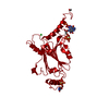
 Assembly


 Spodoptera frugiperda (fall armyworm) / References: UniProt: P11041


 Mass: 35.453 Da / Num. of mol.: 1 / Source method: obtained synthetically / Formula: Cl
 Mass: 24.305 Da / Num. of mol.: 2 / Source method: obtained synthetically / Formula: Mg
 Mass: 507.181 Da / Num. of mol.: 1 / Source method: obtained synthetically / Formula: C10H16N5O13P3 / Comment: ATP, energy-carrying molecule*YM
 Mass: 483.156 Da / Num. of mol.: 1 / Source method: obtained synthetically / Formula: C9H16N3O14P3
 Mass: 62.068 Da / Num. of mol.: 3 / Source method: obtained synthetically / Formula: C2H6O2
 Sample preparation
 / Beamline: BL41XU / Wavelength: 1 Å
 Processing