 Movie
Movie Controller


+ Open data

- Basic information
| Entry | Database: PDB / ID: 5axv | ||||||
|---|---|---|---|---|---|---|---|
| Title | Crystal Structure of Cypovirus Polyhedra R13K Mutant | ||||||
 Components Components | Polyhedrin | ||||||
 Keywords Keywords | STRUCTURAL PROTEIN / INTRACELLULAR CRYSTAL / NUCLEOTIDE BINDING | ||||||
| Function / homology | Cypovirus polyhedrin, Cypovirus 1 type / Cypovirus polyhedrin protein / viral occlusion body / host cell cytoplasm / CYTIDINE-5'-TRIPHOSPHATE / Polyhedrin Function and homology information Function and homology information | ||||||
| Biological species |   Bombyx mori cypovirus 1 Bombyx mori cypovirus 1 | ||||||
| Method |  X-RAY DIFFRACTION / SYNCHROTRON / MOLECULAR REPLACEMENT / Resolution: 2.04 Å X-RAY DIFFRACTION / SYNCHROTRON / MOLECULAR REPLACEMENT / Resolution: 2.04 Å | ||||||
 Authors Authors | Abe, S. / Ijiri, H. / Negishi, H. / Yamanaka, H. / Sasaki, K. / Hirata, K. / Mori, H. / Ueno, T. | ||||||
 Citation Citation | Journal: Adv. Mater. Weinheim / Year: 2015 Title: Design of Enzyme-Encapsulated Protein Containers by In Vivo Crystal Engineering Authors: Abe, S. / Ijiri, H. / Negishi, H. / Yamanaka, H. / Sasaki, K. / Hirata, K. / Mori, H. / Ueno, T. | ||||||
| History |
|
- Structure visualization
| Structure viewer | Molecule: MolmilJmol/JSmol |
|---|
- Downloads & links
-Download
| PDBx/mmCIF format | 5axv.cif.gz | 68.5 KB | Display | PDBx/mmCIF format |
|---|---|---|---|---|
| PDB format | pdb5axv.ent.gz | 48.8 KB | Display | PDB format |
| PDBx/mmJSON format | 5axv.json.gz | Tree view | PDBx/mmJSON format | |
| Others |  Other downloads Other downloads |
-Validation report
| Arichive directory | https://data.pdbj.org/pub/pdb/validation_reports/ax/5axvftp://data.pdbj.org/pub/pdb/validation_reports/ax/5axv | HTTPS FTP |
|---|
-Related structure data
| Related structure data |  5axuC 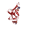 2oh6S C: citing same article ( S: Starting model for refinement |
|---|---|
| Similar structure data |


-Links
 PDBj
PDBj
- Assembly
| Deposited unit | 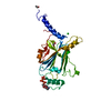
| ||||||||
|---|---|---|---|---|---|---|---|---|---|
| 1 | x 12
| ||||||||
| Unit cell |
|
-Components
| #1: Protein | Mass: 28359.301 Da / Num. of mol.: 1 / Mutation: R13K Source method: isolated from a genetically manipulated source Source: (gene. exp.) Bombyx mori cypovirus 1 / Production host:   Spodoptera frugiperda (fall armyworm) / References: UniProt: P11041 Spodoptera frugiperda (fall armyworm) / References: UniProt: P11041 | ||||
|---|---|---|---|---|---|
| #2: Chemical | ChemComp-CTP /   Mass: 483.156 Da / Num. of mol.: 1 / Source method: obtained synthetically / Formula: C9H16N3O14P3 Mass: 483.156 Da / Num. of mol.: 1 / Source method: obtained synthetically / Formula: C9H16N3O14P3 | ||||
| #3: Chemical | ChemComp-CL /   Mass: 35.453 Da / Num. of mol.: 1 / Source method: obtained synthetically / Formula: Cl Mass: 35.453 Da / Num. of mol.: 1 / Source method: obtained synthetically / Formula: Cl | ||||
| #4: Chemical |   Mass: 62.068 Da / Num. of mol.: 3 / Source method: obtained synthetically / Formula: C2H6O2 Mass: 62.068 Da / Num. of mol.: 3 / Source method: obtained synthetically / Formula: C2H6O2#5: Water | ChemComp-HOH / |  Mass: 18.015 Da / Num. of mol.: 122 / Source method: isolated from a natural source / Formula: H2O Mass: 18.015 Da / Num. of mol.: 122 / Source method: isolated from a natural source / Formula: H2OHas protein modification | Y | |
-Experimental details
-Experiment
| Experiment | Method: X-RAY DIFFRACTION / Number of used crystals: 1 |
|---|
- Sample preparation
| Crystal | Density Matthews: 1.62 Å3/Da / Density % sol: 24.23 % |
|---|---|
| Crystal grow | Temperature: 300 K / Method: in cell / pH: 7 Details: IN VIVO CRYSTALLIZATION IN THE CYTOPLASM OF THE CELL, PH 7.0, TEMPERATURE 300K |
-Data collection
| Diffraction | Mean temperature: 100 K |
|---|---|
| Diffraction source | Source: SYNCHROTRON / Site: SPring-8  / Beamline: BL32XU / Wavelength: 1 Å / Beamline: BL32XU / Wavelength: 1 Å |
| Detector | Type: RAYONIX MX225HE / Detector: CCD / Date: Jul 2, 2011 |
| Radiation | Monochromator: LIQUID NITROGEN COOLED DOUBLE CRYSTAL / Protocol: SINGLE WAVELENGTH / Monochromatic (M) / Laue (L): M / Scattering type: x-ray |
| Radiation wavelength | Wavelength: 1 Å / Relative weight: 1 |
| Reflection | Resolution: 2.04→40 Å / Num. obs: 11308 / % possible obs: 93.3 % / Redundancy: 2.8 % / Biso Wilson estimate: 8.1 Å2 / Rsym value: 0.179 / Net I/σ(I): 6.15 |
| Reflection shell | Resolution: 2.04→2.08 Å / Redundancy: 2.7 % / Mean I/σ(I) obs: 2.2 / Rsym value: 0.48 / % possible all: 97.3 |
- Processing
| Software |
| ||||||||||||||||||||||||||||||||||||||||||||||||||||||||||||||||||||||||||||||||||||||||||||||||||||||||||||||||||||||||||||||||||||||||||||||||||||||||||||||||||||||||||||||||||||||
|---|---|---|---|---|---|---|---|---|---|---|---|---|---|---|---|---|---|---|---|---|---|---|---|---|---|---|---|---|---|---|---|---|---|---|---|---|---|---|---|---|---|---|---|---|---|---|---|---|---|---|---|---|---|---|---|---|---|---|---|---|---|---|---|---|---|---|---|---|---|---|---|---|---|---|---|---|---|---|---|---|---|---|---|---|---|---|---|---|---|---|---|---|---|---|---|---|---|---|---|---|---|---|---|---|---|---|---|---|---|---|---|---|---|---|---|---|---|---|---|---|---|---|---|---|---|---|---|---|---|---|---|---|---|---|---|---|---|---|---|---|---|---|---|---|---|---|---|---|---|---|---|---|---|---|---|---|---|---|---|---|---|---|---|---|---|---|---|---|---|---|---|---|---|---|---|---|---|---|---|---|---|---|---|
| Refinement | Method to determine structure: MOLECULAR REPLACEMENT Starting model: 2OH6 Resolution: 2.04→36.55 Å / Cor.coef. Fo:Fc: 0.953 / Cor.coef. Fo:Fc free: 0.91 / SU B: 4.295 / SU ML: 0.117 / Cross valid method: THROUGHOUT / ESU R: 0.322 / ESU R Free: 0.195 / Stereochemistry target values: MAXIMUM LIKELIHOOD
| ||||||||||||||||||||||||||||||||||||||||||||||||||||||||||||||||||||||||||||||||||||||||||||||||||||||||||||||||||||||||||||||||||||||||||||||||||||||||||||||||||||||||||||||||||||||
| Solvent computation | Ion probe radii: 0.8 Å / Shrinkage radii: 0.8 Å / VDW probe radii: 1.2 Å / Solvent model: MASK | ||||||||||||||||||||||||||||||||||||||||||||||||||||||||||||||||||||||||||||||||||||||||||||||||||||||||||||||||||||||||||||||||||||||||||||||||||||||||||||||||||||||||||||||||||||||
| Displacement parameters | Biso mean: 7.57 Å2 | ||||||||||||||||||||||||||||||||||||||||||||||||||||||||||||||||||||||||||||||||||||||||||||||||||||||||||||||||||||||||||||||||||||||||||||||||||||||||||||||||||||||||||||||||||||||
| Refinement step | Cycle: LAST / Resolution: 2.04→36.55 Å
| ||||||||||||||||||||||||||||||||||||||||||||||||||||||||||||||||||||||||||||||||||||||||||||||||||||||||||||||||||||||||||||||||||||||||||||||||||||||||||||||||||||||||||||||||||||||
| Refine LS restraints |
|