 Movie
Movie Controller

[English] 日本語
 Yorodumi
- PDB-7bbs: Structure of Bg10: an alcohol-tolerant and glucose-stimulated B-g... -
+ Open data

- Basic information
| Entry | Database: PDB / ID: 7bbs | |||||||||
|---|---|---|---|---|---|---|---|---|---|---|
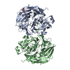
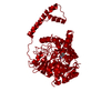
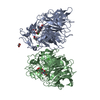
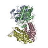
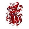
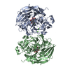
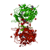
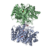
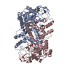
| Title | Structure of Bg10: an alcohol-tolerant and glucose-stimulated B-glucosidase | |||||||||
 Components Components | Beta-glucosidase Bg10 | |||||||||
 Keywords Keywords | HYDROLASE / Glucose-stimulated / GH1 family / metagenomics | |||||||||
| Function / homology |  Function and homology information Function and homology information: / beta-glucosidase / beta-glucosidase activity / carbohydrate metabolic process Similarity search - Function | |||||||||
| Biological species |  uncultured bacterium (environmental samples) uncultured bacterium (environmental samples) | |||||||||
| Method |  X-RAY DIFFRACTION / SYNCHROTRON / MOLECULAR REPLACEMENT / Resolution: 2.3 Å X-RAY DIFFRACTION / SYNCHROTRON / MOLECULAR REPLACEMENT / Resolution: 2.3 Å | |||||||||
 Authors Authors | Maldaner Pereira, P.A. / Gomes-Pepe, E.S. / Silva, S.T.N. / Matias, P.M. / Lemos, E.G.M. | |||||||||
| Funding support |  Brazil, Brazil,  Portugal, 2items Portugal, 2items
| |||||||||
 Citation Citation | Journal: TO BE PUBLISHED Title: Structure of Bg10: an alcohol-tolerant and glucose-stimulated beta-glucosidase Authors: Maldaner Pereira, P.A. / Gomes-Pepe, E.S. / Silva, S.T.N. / Matias, P.M. / Lemos, E.G.M. | |||||||||
| History |
|
- Structure visualization
| Structure viewer | Molecule: MolmilJmol/JSmol |
|---|
- Downloads & links
-Download
| PDBx/mmCIF format | 7bbs.cif.gz | 420.5 KB | Display | PDBx/mmCIF format |
|---|---|---|---|---|
| PDB format | pdb7bbs.ent.gz | 288.1 KB | Display | PDB format |
| PDBx/mmJSON format | 7bbs.json.gz | Tree view | PDBx/mmJSON format | |
| Others |  Other downloads Other downloads |
-Validation report
| Arichive directory | https://data.pdbj.org/pub/pdb/validation_reports/bb/7bbsftp://data.pdbj.org/pub/pdb/validation_reports/bb/7bbs | HTTPS FTP |
|---|
-Related structure data
| Related structure data |  1gnxS S: Starting model for refinement |
|---|---|
| Similar structure data | |
| Experimental dataset #1 | Data reference: 10.5281/zenodo.4327001 / Data set type: diffraction image data |

-Links
 PDBj
PDBj- Assembly
| Deposited unit | 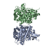
| |||||||||||||||||||||||||||||||||||||
|---|---|---|---|---|---|---|---|---|---|---|---|---|---|---|---|---|---|---|---|---|---|---|---|---|---|---|---|---|---|---|---|---|---|---|---|---|---|---|
| 1 |
| |||||||||||||||||||||||||||||||||||||
| Unit cell |
| |||||||||||||||||||||||||||||||||||||
| Components on special symmetry positions |
| |||||||||||||||||||||||||||||||||||||
| Noncrystallographic symmetry (NCS) | NCS domain:
NCS domain segments:
|
-Components
| #1: Protein | Mass: 54233.020 Da / Num. of mol.: 2 Source method: isolated from a genetically manipulated source Source: (gene. exp.) uncultured bacterium (environmental samples)Production host: #2: Water | ChemComp-HOH / |  Mass: 18.015 Da / Num. of mol.: 21 / Source method: isolated from a natural source / Formula: H2O Mass: 18.015 Da / Num. of mol.: 21 / Source method: isolated from a natural source / Formula: H2O |
|---|
-Experimental details
-Experiment
| Experiment | Method: X-RAY DIFFRACTION / Number of used crystals: 1 |
|---|
- Sample preparation
| Crystal | Density Matthews: 3.32 Å3/Da / Density % sol: 62.9 % / Description: diamond-shaped plates |
|---|---|
| Crystal grow | Temperature: 293 K / Method: vapor diffusion, sitting drop / pH: 7 Details: 12% (v/v) PEG 3550, 250 mM potassium citrate, 4% (v/v) 1,5-pentanediol |
-Data collection
| Diffraction | Mean temperature: 100 K / Serial crystal experiment: N |
|---|---|
| Diffraction source | Source: SYNCHROTRON / Site: Diamond  / Beamline: I04 / Wavelength: 0.9795 Å / Beamline: I04 / Wavelength: 0.9795 Å |
| Detector | Type: DECTRIS EIGER2 XE 16M / Detector: PIXEL / Date: Apr 13, 2019 |
| Radiation | Monochromator: Si 111 double crystal / Protocol: SINGLE WAVELENGTH / Monochromatic (M) / Laue (L): M / Scattering type: x-ray |
| Radiation wavelength | Wavelength: 0.9795 Å / Relative weight: 1 |
| Reflection | Resolution: 2.3→112.13 Å / Num. obs: 33702 / % possible obs: 54.4 % / Redundancy: 3.4 % / Biso Wilson estimate: 27.94 Å2 / CC1/2: 0.982 / Rmerge(I) obs: 0.193 / Rpim(I) all: 0.124 / Rrim(I) all: 0.23 / Net I/σ(I): 5.6 |
| Reflection shell | Resolution: 2.3→2.604 Å / Rmerge(I) obs: 0.643 / Mean I/σ(I) obs: 1.5 / Num. unique obs: 1686 / CC1/2: 0.584 / Rpim(I) all: 0.486 / Rrim(I) all: 0.809 / % possible all: 8.9 |
- Processing
| Software |
| |||||||||||||||||||||||||||||||||||||||||||||||||||||||||||||||||||||||||||||||||||||||||||
|---|---|---|---|---|---|---|---|---|---|---|---|---|---|---|---|---|---|---|---|---|---|---|---|---|---|---|---|---|---|---|---|---|---|---|---|---|---|---|---|---|---|---|---|---|---|---|---|---|---|---|---|---|---|---|---|---|---|---|---|---|---|---|---|---|---|---|---|---|---|---|---|---|---|---|---|---|---|---|---|---|---|---|---|---|---|---|---|---|---|---|---|---|
| Refinement | Method to determine structure: MOLECULAR REPLACEMENT Starting model: 1GNX Resolution: 2.3→112.13 Å / SU ML: 0.336 / Cross valid method: FREE R-VALUE / σ(F): 1.37 / Phase error: 31.498 Stereochemistry target values: GeoStd + Monomer Library + CDL v1.2
| |||||||||||||||||||||||||||||||||||||||||||||||||||||||||||||||||||||||||||||||||||||||||||
| Solvent computation | Shrinkage radii: 0.9 Å / VDW probe radii: 1.11 Å / Solvent model: FLAT BULK SOLVENT MODEL | |||||||||||||||||||||||||||||||||||||||||||||||||||||||||||||||||||||||||||||||||||||||||||
| Displacement parameters | Biso mean: 28.35 Å2 | |||||||||||||||||||||||||||||||||||||||||||||||||||||||||||||||||||||||||||||||||||||||||||
| Refinement step | Cycle: LAST / Resolution: 2.3→112.13 Å
| |||||||||||||||||||||||||||||||||||||||||||||||||||||||||||||||||||||||||||||||||||||||||||
| Refine LS restraints |
| |||||||||||||||||||||||||||||||||||||||||||||||||||||||||||||||||||||||||||||||||||||||||||
| Refine LS restraints NCS | Type: Torsion NCS / Rms dev position: 1.42353283856 Å | |||||||||||||||||||||||||||||||||||||||||||||||||||||||||||||||||||||||||||||||||||||||||||
| LS refinement shell |
| |||||||||||||||||||||||||||||||||||||||||||||||||||||||||||||||||||||||||||||||||||||||||||
| Refinement TLS params. | Method: refined / Refine-ID: X-RAY DIFFRACTION
| |||||||||||||||||||||||||||||||||||||||||||||||||||||||||||||||||||||||||||||||||||||||||||
| Refinement TLS group | Refine-ID: X-RAY DIFFRACTION / Auth seq-ID: 17 - 484
|
