 Movie
Movie Controller

[English] 日本語
 Yorodumi
- PDB-1cq1: Soluble Quinoprotein Glucose Dehydrogenase from Acinetobacter Cal... -
+ Open data

- Basic information
| Entry | Database: PDB / ID: 1cq1 | ||||||
|---|---|---|---|---|---|---|---|
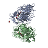
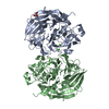
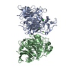
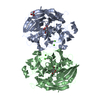
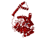
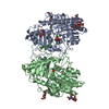
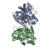
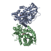
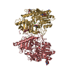
| Title | Soluble Quinoprotein Glucose Dehydrogenase from Acinetobacter Calcoaceticus in Complex with PQQH2 and Glucose | ||||||
 Components Components | SOLUBLE QUINOPROTEIN GLUCOSE DEHYDROGENASE | ||||||
 Keywords Keywords | OXIDOREDUCTASE / BETA-PROPELLER / SUPERBARREL / COMPLEX WITH COFACTOR AND SUBSTRATE | ||||||
| Function / homology |  Function and homology information Function and homology informationglucose 1-dehydrogenase (PQQ, quinone) / quinoprotein glucose dehydrogenase activity / metal ion binding Similarity search - Function | ||||||
| Biological species |  Acinetobacter calcoaceticus (bacteria) Acinetobacter calcoaceticus (bacteria) | ||||||
| Method |  X-RAY DIFFRACTION / SYNCHROTRON / Resolution: 1.9 Å X-RAY DIFFRACTION / SYNCHROTRON / Resolution: 1.9 Å | ||||||
 Authors Authors | Oubrie, A. / Rozeboom, H.J. / Dijkstra, B.W. | ||||||
 Citation Citation | Journal: EMBO J. / Year: 1999 Title: Structure and mechanism of soluble quinoprotein glucose dehydrogenase. Authors: Oubrie, A. / Rozeboom, H.J. / Kalk, K.H. / Olsthoorn, A.J. / Duine, J.A. / Dijkstra, B.W. #1: Journal: J.Mol.Biol. / Year: 1999Title: The 1.7 Angstrom crystal structure of the apo form of the soluble quinoprotein glucose dehydrogenase from Acinetobacter calcoaceticus reveals a novel internal sequence repeat Authors: Oubrie, A. / Rozeboom, H.J. / Kalk, K.H. / Duine, J.A. / Dijkstra, B.W. | ||||||
| History |
|
- Structure visualization
| Structure viewer | Molecule: MolmilJmol/JSmol |
|---|
- Downloads & links
-Download
| PDBx/mmCIF format | 1cq1.cif.gz | 199.4 KB | Display | PDBx/mmCIF format |
|---|---|---|---|---|
| PDB format | pdb1cq1.ent.gz | 158.1 KB | Display | PDB format |
| PDBx/mmJSON format | 1cq1.json.gz | Tree view | PDBx/mmJSON format | |
| Others |  Other downloads Other downloads |
-Validation report
| Arichive directory | https://data.pdbj.org/pub/pdb/validation_reports/cq/1cq1ftp://data.pdbj.org/pub/pdb/validation_reports/cq/1cq1 | HTTPS FTP |
|---|
-Related structure data


-Links
 PDBj
PDBj


- Assembly
| Deposited unit | 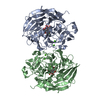
| ||||||||
|---|---|---|---|---|---|---|---|---|---|
| 1 |
| ||||||||
| Unit cell |
|
-Components
| #1: Protein | Mass: 50293.207 Da / Num. of mol.: 2 Source method: isolated from a genetically manipulated source Source: (gene. exp.) Acinetobacter calcoaceticus (bacteria) / Cellular location: PERIPLASM / Production host: #2: Sugar |   Type: D-saccharide, beta linking / Mass: 180.156 Da / Num. of mol.: 2 / Source method: obtained synthetically / Formula: C6H12O6 Type: D-saccharide, beta linking / Mass: 180.156 Da / Num. of mol.: 2 / Source method: obtained synthetically / Formula: C6H12O6#3: Chemical | ChemComp-CA /   Mass: 40.078 Da / Num. of mol.: 6 / Source method: obtained synthetically / Formula: Ca Mass: 40.078 Da / Num. of mol.: 6 / Source method: obtained synthetically / Formula: Ca#4: Chemical |   Mass: 330.206 Da / Num. of mol.: 2 / Source method: obtained synthetically / Formula: C14H6N2O8 Mass: 330.206 Da / Num. of mol.: 2 / Source method: obtained synthetically / Formula: C14H6N2O8#5: Water | ChemComp-HOH / |  Mass: 18.015 Da / Num. of mol.: 590 / Source method: isolated from a natural source / Formula: H2O Mass: 18.015 Da / Num. of mol.: 590 / Source method: isolated from a natural source / Formula: H2OHas protein modification | Y | |
|---|
-Experimental details
-Experiment
| Experiment | Method: X-RAY DIFFRACTION / Number of used crystals: 1 |
|---|
- Sample preparation
| Crystal | Density Matthews: 2.31 Å3/Da / Density % sol: 46.72 % | ||||||||||||||||||||||||||||||
|---|---|---|---|---|---|---|---|---|---|---|---|---|---|---|---|---|---|---|---|---|---|---|---|---|---|---|---|---|---|---|---|
| Crystal grow | Temperature: 293 K / Method: vapor diffusion, hanging drop / pH: 9.2 Details: PEG 6000, SODIUM CHLORIDE, CALCIUM CHLORIDE, TRIS, GLYCINE, pH 9.2, VAPOR DIFFUSION, HANGING DROP, temperature 293K | ||||||||||||||||||||||||||||||
| Crystal grow | *PLUS | ||||||||||||||||||||||||||||||
| Components of the solutions | *PLUS
|
-Data collection
| Diffraction | Mean temperature: 100 K |
|---|---|
| Diffraction source | Source: SYNCHROTRON / Site: EMBL/DESY, HAMBURG  / Beamline: BW7A / Wavelength: 1.0736 / Beamline: BW7A / Wavelength: 1.0736 |
| Detector | Type: MARRESEARCH / Detector: IMAGE PLATE / Date: Sep 23, 1998 |
| Radiation | Protocol: SINGLE WAVELENGTH / Monochromatic (M) / Laue (L): M / Scattering type: x-ray |
| Radiation wavelength | Wavelength: 1.0736 Å / Relative weight: 1 |
| Reflection | Resolution: 1.75→50 Å / Num. all: 92216 / Num. obs: 92032 / % possible obs: 99.8 % / Observed criterion σ(F): 0 / Observed criterion σ(I): 0 / Redundancy: 3.5 % / Biso Wilson estimate: 13.6 Å2 / Rmerge(I) obs: 0.064 / Net I/σ(I): 18.6 |
| Reflection shell | Resolution: 1.75→1.78 Å / Redundancy: 3.3 % / Rmerge(I) obs: 0.814 / % possible all: 98.1 |
| Reflection shell | *PLUS % possible obs: 98.1 % |
- Processing
| Software |
| ||||||||||||||||||||||||||||||||||||||||||||||||||||||||||||
|---|---|---|---|---|---|---|---|---|---|---|---|---|---|---|---|---|---|---|---|---|---|---|---|---|---|---|---|---|---|---|---|---|---|---|---|---|---|---|---|---|---|---|---|---|---|---|---|---|---|---|---|---|---|---|---|---|---|---|---|---|---|
| Refinement | Resolution: 1.9→20 Å / σ(F): 0 / σ(I): 0 / Stereochemistry target values: ENGH & HUBER
| ||||||||||||||||||||||||||||||||||||||||||||||||||||||||||||
| Refinement step | Cycle: LAST / Resolution: 1.9→20 Å
| ||||||||||||||||||||||||||||||||||||||||||||||||||||||||||||
| Refine LS restraints |
| ||||||||||||||||||||||||||||||||||||||||||||||||||||||||||||
| Software | *PLUS Name: 'REFMAC, X-PLOR 3.843' / Classification: refinement | ||||||||||||||||||||||||||||||||||||||||||||||||||||||||||||
| Refine LS restraints | *PLUS Type: x_angle_deg / Dev ideal: 3 |
