 ムービー
ムービー コントローラー


+ データを開く

- 基本情報
基本情報
| 登録情報 | データベース: PDB / ID: 7bbc | ||||||
|---|---|---|---|---|---|---|---|
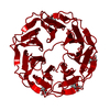
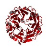
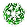
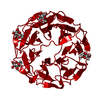
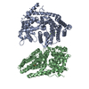
| タイトル | Joint X-ray/neutron room temperature structure of perdeuterated PLL lectin in complex with perdeuterated L-fucose | ||||||
 要素 要素 | PLL lectin | ||||||
 キーワード キーワード | SUGAR BINDING PROTEIN / Complex / propeller | ||||||
| 機能・相同性 | Protein of unknown function DUF346 / Repeat of unknown function (DUF346) / alpha-L-fucopyranose / beta-L-fucopyranose / Uncharacterized protein 機能・相同性情報 機能・相同性情報 | ||||||
| 生物種 |  Photorhabdus laumondii (バクテリア) Photorhabdus laumondii (バクテリア) | ||||||
| 手法 |  X線回折 / 中性子回折 / NUCLEAR REACTOR / 分子置換 / 解像度: 1.84 Å X線回折 / 中性子回折 / NUCLEAR REACTOR / 分子置換 / 解像度: 1.84 Å | ||||||
 データ登録者 データ登録者 | Gajdos, L. / Blakeley, M.P. / Kumar, A. / Wimmerova, M. / Haertlein, M. / Forsyth, V.T. / Imberty, A. / Devos, J.M. | ||||||
| 資金援助 |  フランス, 1件 フランス, 1件
| ||||||
 引用 引用 | ジャーナル: Structure / 年: 2021 タイトル: Visualization of hydrogen atoms in a perdeuterated lectin-fucose complex reveals key details of protein-carbohydrate interactions. 著者: Gajdos, L. / Blakeley, M.P. / Kumar, A. / Wimmerova, M. / Haertlein, M. / Forsyth, V.T. / Imberty, A. / Devos, J.M. | ||||||
| 履歴 |
|
- 構造の表示
構造の表示
| 構造ビューア | 分子: MolmilJmol/JSmol |
|---|
- ダウンロードとリンク
-ダウンロード
| PDBx/mmCIF形式 | 7bbc.cif.gz | 209.4 KB | 表示 | PDBx/mmCIF形式 |
|---|---|---|---|---|
| PDB形式 | pdb7bbc.ent.gz | 139.8 KB | 表示 | PDB形式 |
| PDBx/mmJSON形式 | 7bbc.json.gz | ツリー表示 | PDBx/mmJSON形式 | |
| その他 |  その他のダウンロード その他のダウンロード |
-検証レポート
| アーカイブディレクトリ | https://data.pdbj.org/pub/pdb/validation_reports/bb/7bbcftp://data.pdbj.org/pub/pdb/validation_reports/bb/7bbc | HTTPS FTP |
|---|
-関連構造データ


-リンク
 PDBj
PDBj
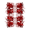
- 集合体
集合体
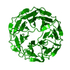
| 登録構造単位 | 
| ||||||||||||
|---|---|---|---|---|---|---|---|---|---|---|---|---|---|
| 1 | 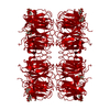
| ||||||||||||
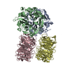
| 単位格子 |
| ||||||||||||
| Components on special symmetry positions |
|
-要素
| #1: タンパク質 | 分子量: 41944.949 Da / 分子数: 1 / 由来タイプ: 組換発現 由来: (組換発現) Photorhabdus laumondii (バクテリア)遺伝子: CKY10_20885 / 発現宿主: | ||||||||
|---|---|---|---|---|---|---|---|---|---|
| #2: 糖 |   タイプ: L-saccharide, beta linking / 分子量: 164.156 Da / 分子数: 3 / 由来タイプ: 合成 / 式: C6H12O5 / タイプ: SUBJECT OF INVESTIGATION タイプ: L-saccharide, beta linking / 分子量: 164.156 Da / 分子数: 3 / 由来タイプ: 合成 / 式: C6H12O5 / タイプ: SUBJECT OF INVESTIGATION#3: 糖 |   タイプ: L-saccharide, alpha linking / 分子量: 164.156 Da / 分子数: 3 / 由来タイプ: 合成 / 式: C6H12O5 / タイプ: SUBJECT OF INVESTIGATION タイプ: L-saccharide, alpha linking / 分子量: 164.156 Da / 分子数: 3 / 由来タイプ: 合成 / 式: C6H12O5 / タイプ: SUBJECT OF INVESTIGATION#4: 水 | ChemComp-HOH / |  分子量: 18.015 Da / 分子数: 334 / 由来タイプ: 天然 / 式: H2O 分子量: 18.015 Da / 分子数: 334 / 由来タイプ: 天然 / 式: H2O研究の焦点であるリガンドがあるか | Y | Has protein modification | Y | |
-実験情報
-実験
| 実験 |
|
|---|
- 試料調製
試料調製
| 結晶 | マシュー密度: 3.08 Å3/Da / 溶媒含有率: 60.01 % |
|---|---|
| 結晶化 | 温度: 293 K / 手法: 蒸気拡散法, シッティングドロップ法 詳細: 0.1 M Na/K tartrate dissolved in D2O, soaked with 50 mM perdeuterated fucose-d12 dissolved in D2O |
-データ収集
| 回折 |
| ||||||||||||||||||||||||
|---|---|---|---|---|---|---|---|---|---|---|---|---|---|---|---|---|---|---|---|---|---|---|---|---|---|
| 放射光源 |
| ||||||||||||||||||||||||
| 検出器 |
| ||||||||||||||||||||||||
| 放射 |
| ||||||||||||||||||||||||
| 放射波長 |
| ||||||||||||||||||||||||
| 反射 | Biso Wilson estimate: 19.08 Å2 / Entry-ID: 7BBC
| ||||||||||||||||||||||||
| 反射 シェル |
|
- 解析
解析
| ソフトウェア |
| |||||||||||||||||||||||||||||||||||||||||||||||||||||||||||||||||||||||||||||||||||||||||||||||||||||||||||||||||||||||
|---|---|---|---|---|---|---|---|---|---|---|---|---|---|---|---|---|---|---|---|---|---|---|---|---|---|---|---|---|---|---|---|---|---|---|---|---|---|---|---|---|---|---|---|---|---|---|---|---|---|---|---|---|---|---|---|---|---|---|---|---|---|---|---|---|---|---|---|---|---|---|---|---|---|---|---|---|---|---|---|---|---|---|---|---|---|---|---|---|---|---|---|---|---|---|---|---|---|---|---|---|---|---|---|---|---|---|---|---|---|---|---|---|---|---|---|---|---|---|---|---|
| 精密化 | Biso mean: 34.65 Å2 / % reflection Rfree: 5 % / SU ML: 0.1415 / 交差検証法: FREE R-VALUE / 構造決定の手法:
| |||||||||||||||||||||||||||||||||||||||||||||||||||||||||||||||||||||||||||||||||||||||||||||||||||||||||||||||||||||||
| 精密化ステップ | サイクル: LAST / 解像度: 1.84→33 Å
| |||||||||||||||||||||||||||||||||||||||||||||||||||||||||||||||||||||||||||||||||||||||||||||||||||||||||||||||||||||||
| 拘束条件 |
| |||||||||||||||||||||||||||||||||||||||||||||||||||||||||||||||||||||||||||||||||||||||||||||||||||||||||||||||||||||||
| LS精密化 シェル |
|
