 Movie
Movie Controller

[English] 日本語
 Yorodumi
- PDB-7b7f: Room temperature X-ray structure of H/D-exchanged PLL lectin in c... -
+ Open data

- Basic information
| Entry | Database: PDB / ID: 7b7f | ||||||
|---|---|---|---|---|---|---|---|
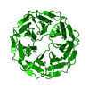
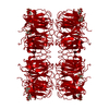
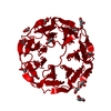
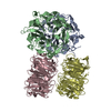
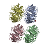
| Title | Room temperature X-ray structure of H/D-exchanged PLL lectin in complex with L-fucose | ||||||
 Components Components | PLL lectin | ||||||
 Keywords Keywords | SUGAR BINDING PROTEIN / Complex / propeller | ||||||
| Function / homology | Protein of unknown function DUF346 / Repeat of unknown function (DUF346) / alpha-L-fucopyranose / beta-L-fucopyranose / Uncharacterized protein Function and homology information Function and homology information | ||||||
| Biological species |  Photorhabdus laumondii (bacteria) Photorhabdus laumondii (bacteria) | ||||||
| Method |  X-RAY DIFFRACTION / SYNCHROTRON / MOLECULAR REPLACEMENT / Resolution: 1.6 Å X-RAY DIFFRACTION / SYNCHROTRON / MOLECULAR REPLACEMENT / Resolution: 1.6 Å | ||||||
 Authors Authors | Gajdos, L. / Blakeley, M.P. / Kumar, A. / Wimmerova, M. / Haertlein, M. / Forsyth, V.T. / Imberty, A. / Devos, J.M. | ||||||
| Funding support |  France, 1items France, 1items
| ||||||
 Citation Citation | Journal: Structure / Year: 2021 Title: Visualization of hydrogen atoms in a perdeuterated lectin-fucose complex reveals key details of protein-carbohydrate interactions. Authors: Gajdos, L. / Blakeley, M.P. / Kumar, A. / Wimmerova, M. / Haertlein, M. / Forsyth, V.T. / Imberty, A. / Devos, J.M. | ||||||
| History |
|
- Structure visualization
| Structure viewer | Molecule: MolmilJmol/JSmol |
|---|
- Downloads & links
-Download
| PDBx/mmCIF format | 7b7f.cif.gz | 123.8 KB | Display | PDBx/mmCIF format |
|---|---|---|---|---|
| PDB format | pdb7b7f.ent.gz | 77.1 KB | Display | PDB format |
| PDBx/mmJSON format | 7b7f.json.gz | Tree view | PDBx/mmJSON format | |
| Others |  Other downloads Other downloads |
-Validation report
| Arichive directory | https://data.pdbj.org/pub/pdb/validation_reports/b7/7b7fftp://data.pdbj.org/pub/pdb/validation_reports/b7/7b7f | HTTPS FTP |
|---|
-Related structure data
| Related structure data | 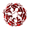 7b7cC  7b7eC  7bb4C 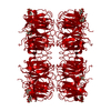 7bbcC  7bbiC  5c9oS S: Starting model for refinement C: citing same article ( |
|---|---|
| Similar structure data |


-Links
 PDBj
PDBj
- Assembly
| Deposited unit | 
| ||||||||||||
|---|---|---|---|---|---|---|---|---|---|---|---|---|---|
| 1 | 
| ||||||||||||
| Unit cell |
| ||||||||||||
| Components on special symmetry positions |
|
-Components
| #1: Protein | Mass: 41944.949 Da / Num. of mol.: 1 Source method: isolated from a genetically manipulated source Source: (gene. exp.) Photorhabdus laumondii (bacteria) / Gene: CKY10_20885 / Production host: | ||||||||
|---|---|---|---|---|---|---|---|---|---|
| #2: Sugar | ChemComp-FUC /   Type: L-saccharide, alpha linking / Mass: 164.156 Da / Num. of mol.: 4 / Source method: obtained synthetically / Formula: C6H12O5 / Feature type: SUBJECT OF INVESTIGATION Type: L-saccharide, alpha linking / Mass: 164.156 Da / Num. of mol.: 4 / Source method: obtained synthetically / Formula: C6H12O5 / Feature type: SUBJECT OF INVESTIGATION#3: Sugar | ChemComp-FUL /   Type: L-saccharide, beta linking / Mass: 164.156 Da / Num. of mol.: 4 / Source method: obtained synthetically / Formula: C6H12O5 / Feature type: SUBJECT OF INVESTIGATION Type: L-saccharide, beta linking / Mass: 164.156 Da / Num. of mol.: 4 / Source method: obtained synthetically / Formula: C6H12O5 / Feature type: SUBJECT OF INVESTIGATION#4: Water | ChemComp-HOH / |  Mass: 18.015 Da / Num. of mol.: 406 / Source method: isolated from a natural source / Formula: H2O Mass: 18.015 Da / Num. of mol.: 406 / Source method: isolated from a natural source / Formula: H2OHas ligand of interest | Y | Has protein modification | Y | |
-Experimental details
-Experiment
| Experiment | Method: X-RAY DIFFRACTION / Number of used crystals: 1 |
|---|
- Sample preparation
| Crystal | Density Matthews: 3.08 Å3/Da / Density % sol: 60 % |
|---|---|
| Crystal grow | Temperature: 293 K / Method: vapor diffusion, sitting drop Details: 0.4 M Na/K tartrate dissolved in D2O, soaked with 50 mM L-fucose dissolved in D2O |
-Data collection
| Diffraction | Mean temperature: 293 K / Serial crystal experiment: N |
|---|---|
| Diffraction source | Source: SYNCHROTRON / Site: ESRF / Beamline: BM30A / Wavelength: 0.9796 Å |
| Detector | Type: ADSC QUANTUM 315r / Detector: CCD / Date: Oct 24, 2018 |
| Radiation | Protocol: SINGLE WAVELENGTH / Monochromatic (M) / Laue (L): M / Scattering type: x-ray |
| Radiation wavelength | Wavelength: 0.9796 Å / Relative weight: 1 |
| Reflection | Resolution: 1.6→46 Å / Num. obs: 67576 / % possible obs: 98.7 % / Redundancy: 5.2 % / Biso Wilson estimate: 19.77 Å2 / CC1/2: 1 / Rmerge(I) obs: 0.06 / Net I/σ(I): 16.2 |
| Reflection shell | Resolution: 1.6→1.64 Å / Num. unique obs: 4953 / CC1/2: 0.7 |
- Processing
| Software |
| |||||||||||||||||||||||||||||||||||||||||||||||||||||||||||||||||||||||||||||||||||||||||||||||||||||||||||||||||||||||||||||||||||||||||||||||||||||||||||||||||||||||||||||||
|---|---|---|---|---|---|---|---|---|---|---|---|---|---|---|---|---|---|---|---|---|---|---|---|---|---|---|---|---|---|---|---|---|---|---|---|---|---|---|---|---|---|---|---|---|---|---|---|---|---|---|---|---|---|---|---|---|---|---|---|---|---|---|---|---|---|---|---|---|---|---|---|---|---|---|---|---|---|---|---|---|---|---|---|---|---|---|---|---|---|---|---|---|---|---|---|---|---|---|---|---|---|---|---|---|---|---|---|---|---|---|---|---|---|---|---|---|---|---|---|---|---|---|---|---|---|---|---|---|---|---|---|---|---|---|---|---|---|---|---|---|---|---|---|---|---|---|---|---|---|---|---|---|---|---|---|---|---|---|---|---|---|---|---|---|---|---|---|---|---|---|---|---|---|---|---|---|
| Refinement | Method to determine structure: MOLECULAR REPLACEMENT Starting model: 5c9o Resolution: 1.6→39.82 Å / SU ML: 0.2352 / Cross valid method: FREE R-VALUE / σ(F): 1.35 / Phase error: 17.2839 Stereochemistry target values: GeoStd + Monomer Library + CDL v1.2
| |||||||||||||||||||||||||||||||||||||||||||||||||||||||||||||||||||||||||||||||||||||||||||||||||||||||||||||||||||||||||||||||||||||||||||||||||||||||||||||||||||||||||||||||
| Solvent computation | Shrinkage radii: 0.9 Å / VDW probe radii: 1.11 Å / Solvent model: FLAT BULK SOLVENT MODEL | |||||||||||||||||||||||||||||||||||||||||||||||||||||||||||||||||||||||||||||||||||||||||||||||||||||||||||||||||||||||||||||||||||||||||||||||||||||||||||||||||||||||||||||||
| Displacement parameters | Biso mean: 22.55 Å2 | |||||||||||||||||||||||||||||||||||||||||||||||||||||||||||||||||||||||||||||||||||||||||||||||||||||||||||||||||||||||||||||||||||||||||||||||||||||||||||||||||||||||||||||||
| Refinement step | Cycle: LAST / Resolution: 1.6→39.82 Å
| |||||||||||||||||||||||||||||||||||||||||||||||||||||||||||||||||||||||||||||||||||||||||||||||||||||||||||||||||||||||||||||||||||||||||||||||||||||||||||||||||||||||||||||||
| Refine LS restraints |
| |||||||||||||||||||||||||||||||||||||||||||||||||||||||||||||||||||||||||||||||||||||||||||||||||||||||||||||||||||||||||||||||||||||||||||||||||||||||||||||||||||||||||||||||
| LS refinement shell |
|
