 Movie
Movie Controller


+ Open data

- Basic information
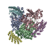
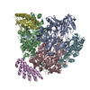
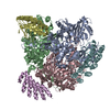
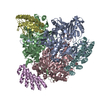
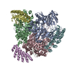
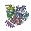
| Entry | Database: PDB / ID: 7b8r | ||||||||||||
|---|---|---|---|---|---|---|---|---|---|---|---|---|---|
| Title | Doxycycline bound structure of bacterial efflux pump. | ||||||||||||
 Components Components |
| ||||||||||||
 Keywords Keywords | TRANSPORT PROTEIN / RND transporter / multidrug resistance / efflux pump / antibiotics | ||||||||||||
| Function / homology |  Function and homology information Function and homology informationalkane transmembrane transporter activity / alkane transport / enterobactin transport / enterobactin transmembrane transporter activity / xenobiotic detoxification by transmembrane export across the cell outer membrane / periplasmic side of plasma membrane / efflux pump complex / Iron assimilation using enterobactin / bile acid transmembrane transporter activity / Antimicrobial resistance ...alkane transmembrane transporter activity / alkane transport / enterobactin transport / enterobactin transmembrane transporter activity / xenobiotic detoxification by transmembrane export across the cell outer membrane / periplasmic side of plasma membrane / efflux pump complex / Iron assimilation using enterobactin / bile acid transmembrane transporter activity / Antimicrobial resistance / bile acid and bile salt transport / Secretion of toxins / xenobiotic transmembrane transporter activity / efflux transmembrane transporter activity / fatty acid transport / xenobiotic transport / response to toxic substance / response to xenobiotic stimulus / response to antibiotic / membrane / identical protein binding / plasma membrane Similarity search - Function | ||||||||||||
| Biological species |  synthetic construct (others) | ||||||||||||
| Method |  X-RAY DIFFRACTION / SYNCHROTRON / MOLECULAR REPLACEMENT / Resolution: 2.1 Å X-RAY DIFFRACTION / SYNCHROTRON / MOLECULAR REPLACEMENT / Resolution: 2.1 Å | ||||||||||||
 Authors Authors | Wilhelm, J. / Sjuts, H. / Pos, K.M. | ||||||||||||
| Funding support |  Germany, European Union, 3items Germany, European Union, 3items
| ||||||||||||
 Citation Citation | Journal: Nat Commun / Year: 2021 Title: Structural and functional analysis of the promiscuous AcrB and AdeB efflux pumps suggests different drug binding mechanisms. Authors: Alina Ornik-Cha / Julia Wilhelm / Jessica Kobylka / Hanno Sjuts / Attilio V Vargiu / Giuliano Malloci / Julian Reitz / Anja Seybert / Achilleas S Frangakis / Klaas M Pos /  Abstract: Upon antibiotic stress Gram-negative pathogens deploy resistance-nodulation-cell division-type tripartite efflux pumps. These include a H/drug antiporter module that recognizes structurally diverse ...Upon antibiotic stress Gram-negative pathogens deploy resistance-nodulation-cell division-type tripartite efflux pumps. These include a H/drug antiporter module that recognizes structurally diverse substances, including antibiotics. Here, we show the 3.5 Å structure of subunit AdeB from the Acinetobacter baumannii AdeABC efflux pump solved by single-particle cryo-electron microscopy. The AdeB trimer adopts mainly a resting state with all protomers in a conformation devoid of transport channels or antibiotic binding sites. However, 10% of the protomers adopt a state where three transport channels lead to the closed substrate (deep) binding pocket. A comparison between drug binding of AdeB and Escherichia coli AcrB is made via activity analysis of 20 AdeB variants, selected on basis of side chain interactions with antibiotics observed in the AcrB periplasmic domain X-ray co-structures with fusidic acid (2.3 Å), doxycycline (2.1 Å) and levofloxacin (2.7 Å). AdeABC, compared to AcrAB-TolC, confers higher resistance to E. coli towards polyaromatic compounds and lower resistance towards antibiotic compounds. | ||||||||||||
| History |
|
- Structure visualization
| Structure viewer | Molecule: MolmilJmol/JSmol |
|---|
- Downloads & links
-Download
| PDBx/mmCIF format | 7b8r.cif.gz | 441 KB | Display | PDBx/mmCIF format |
|---|---|---|---|---|
| PDB format | pdb7b8r.ent.gz | 356.1 KB | Display | PDB format |
| PDBx/mmJSON format | 7b8r.json.gz | Tree view | PDBx/mmJSON format | |
| Others |  Other downloads Other downloads |
-Validation report
| Arichive directory | https://data.pdbj.org/pub/pdb/validation_reports/b8/7b8rftp://data.pdbj.org/pub/pdb/validation_reports/b8/7b8r | HTTPS FTP |
|---|
-Related structure data
| Related structure data |  7b8pC  7b8qC  7b8sC  7b8tC  5en5S S: Starting model for refinement C: citing same article ( |
|---|---|
| Similar structure data |


-Links
 PDBj
PDBj
- Assembly
| Deposited unit | 
| ||||||||||
|---|---|---|---|---|---|---|---|---|---|---|---|
| 1 |
| ||||||||||
| Unit cell |
|
-Components
| #1: Protein | Mass: 66099.047 Da / Num. of mol.: 3 Source method: isolated from a genetically manipulated source Source: (gene. exp.) Strain: K12 / Gene: acrB, acrE, b0462, JW0451 / Production host: #2: Protein | Mass: 18317.566 Da / Num. of mol.: 3 Source method: isolated from a genetically manipulated source Source: (gene. exp.) synthetic construct (others) / Production host: #3: Chemical | ChemComp-PEG /   Mass: 106.120 Da / Num. of mol.: 5 / Source method: obtained synthetically / Formula: C4H10O3 Mass: 106.120 Da / Num. of mol.: 5 / Source method: obtained synthetically / Formula: C4H10O3#4: Chemical |   Mass: 444.435 Da / Num. of mol.: 2 / Source method: obtained synthetically / Formula: C22H24N2O8 / Feature type: SUBJECT OF INVESTIGATION / Comment: antibiotic*YM Mass: 444.435 Da / Num. of mol.: 2 / Source method: obtained synthetically / Formula: C22H24N2O8 / Feature type: SUBJECT OF INVESTIGATION / Comment: antibiotic*YM#5: Water | ChemComp-HOH / |  Mass: 18.015 Da / Num. of mol.: 689 / Source method: isolated from a natural source / Formula: H2O Mass: 18.015 Da / Num. of mol.: 689 / Source method: isolated from a natural source / Formula: H2OHas ligand of interest | Y | |
|---|
-Experimental details
-Experiment
| Experiment | Method: X-RAY DIFFRACTION / Number of used crystals: 1 |
|---|
- Sample preparation
| Crystal | Density Matthews: 2.71 Å3/Da / Density % sol: 54.67 % |
|---|---|
| Crystal grow | Temperature: 291 K / Method: vapor diffusion, hanging drop / Details: 0.1 M MES pH 6.5, 0.21 M NaCl, 11.5% PEG4000 |
-Data collection
| Diffraction | Mean temperature: 100 K / Serial crystal experiment: N |
|---|---|
| Diffraction source | Source: SYNCHROTRON / Site: PETRA III, EMBL c/o DESY / Beamline: P13 (MX1) / Wavelength: 0.976253 Å |
| Detector | Type: DECTRIS PILATUS 6M-F / Detector: PIXEL / Date: Sep 12, 2018 |
| Radiation | Protocol: SINGLE WAVELENGTH / Monochromatic (M) / Laue (L): M / Scattering type: x-ray |
| Radiation wavelength | Wavelength: 0.976253 Å / Relative weight: 1 |
| Reflection | Resolution: 2.1→48.799 Å / Num. obs: 158834 / % possible obs: 98.81 % / Redundancy: 13.7 % / Biso Wilson estimate: 41.76 Å2 / CC1/2: 0.999 / Net I/σ(I): 14.87 |
| Reflection shell | Resolution: 2.1→2.175 Å / Redundancy: 14.3 % / Mean I/σ(I) obs: 1.46 / Num. unique obs: 15593 / CC1/2: 0.65 / % possible all: 98.01 |
- Processing
| Software |
| ||||||||||||||||||||||||||||||||||||||||||||||||||||||||||||||||||||||||||||||||||||||||||||||||||||||||||||||||||||||||||||||||||||||||||||||||||||||||||||||||||||||||||||||||||||||||||
|---|---|---|---|---|---|---|---|---|---|---|---|---|---|---|---|---|---|---|---|---|---|---|---|---|---|---|---|---|---|---|---|---|---|---|---|---|---|---|---|---|---|---|---|---|---|---|---|---|---|---|---|---|---|---|---|---|---|---|---|---|---|---|---|---|---|---|---|---|---|---|---|---|---|---|---|---|---|---|---|---|---|---|---|---|---|---|---|---|---|---|---|---|---|---|---|---|---|---|---|---|---|---|---|---|---|---|---|---|---|---|---|---|---|---|---|---|---|---|---|---|---|---|---|---|---|---|---|---|---|---|---|---|---|---|---|---|---|---|---|---|---|---|---|---|---|---|---|---|---|---|---|---|---|---|---|---|---|---|---|---|---|---|---|---|---|---|---|---|---|---|---|---|---|---|---|---|---|---|---|---|---|---|---|---|---|---|---|
| Refinement | Method to determine structure: MOLECULAR REPLACEMENT Starting model: 5EN5 Resolution: 2.1→48.799 Å / SU ML: 0.26 / Cross valid method: THROUGHOUT / σ(F): 1.34 / Phase error: 26.25 / Stereochemistry target values: ML
| ||||||||||||||||||||||||||||||||||||||||||||||||||||||||||||||||||||||||||||||||||||||||||||||||||||||||||||||||||||||||||||||||||||||||||||||||||||||||||||||||||||||||||||||||||||||||||
| Solvent computation | Shrinkage radii: 0.9 Å / VDW probe radii: 1.11 Å / Solvent model: FLAT BULK SOLVENT MODEL | ||||||||||||||||||||||||||||||||||||||||||||||||||||||||||||||||||||||||||||||||||||||||||||||||||||||||||||||||||||||||||||||||||||||||||||||||||||||||||||||||||||||||||||||||||||||||||
| Displacement parameters | Biso max: 111.01 Å2 / Biso mean: 51.4338 Å2 / Biso min: 23.45 Å2 | ||||||||||||||||||||||||||||||||||||||||||||||||||||||||||||||||||||||||||||||||||||||||||||||||||||||||||||||||||||||||||||||||||||||||||||||||||||||||||||||||||||||||||||||||||||||||||
| Refinement step | Cycle: final / Resolution: 2.1→48.799 Å
| ||||||||||||||||||||||||||||||||||||||||||||||||||||||||||||||||||||||||||||||||||||||||||||||||||||||||||||||||||||||||||||||||||||||||||||||||||||||||||||||||||||||||||||||||||||||||||
| LS refinement shell | Refine-ID: X-RAY DIFFRACTION / Rfactor Rfree error: 0
|
