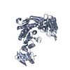
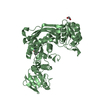
 Movie
Movie Controller


+ Open data

- Basic information
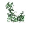
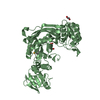
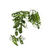
| Entry | Database: PDB / ID: 7b6q | ||||||
|---|---|---|---|---|---|---|---|
| Title | Crystal structure of MurE from E.coli in complex with Z57299526 | ||||||
 Components Components | UDP-N-acetylmuramoyl-L-alanyl-D-glutamate-2,6-diaminopimelate ligase | ||||||
 Keywords Keywords | BIOSYNTHETIC PROTEIN / UDP-N-acetylmuramoyl-L-alanyl-D-glutamate-2 / 6-diaminopimelate ligase cell wall biosynthesis ligase drug target / Structural Genomics / Structural Genomics Consortium / SGC / fragment screening | ||||||
| Function / homology |  Function and homology information Function and homology informationUDP-N-acetylmuramoyl-L-alanyl-D-glutamate-2,6-diaminopimelate ligase / UDP-N-acetylmuramoylalanyl-D-glutamate-2,6-diaminopimelate ligase activity / peptidoglycan biosynthetic process / cell wall organization / regulation of cell shape / cell division / magnesium ion binding / ATP binding / cytosol Similarity search - Function | ||||||
| Biological species |  | ||||||
| Method |  X-RAY DIFFRACTION / SYNCHROTRON / MOLECULAR REPLACEMENT / Resolution: 1.82 Å X-RAY DIFFRACTION / SYNCHROTRON / MOLECULAR REPLACEMENT / Resolution: 1.82 Å | ||||||
 Authors Authors | Koekemoer, L. / Steindel, M. / Fairhead, M. / Talon, R. / Douangamath, A. / Arrowsmith, C.H. / Edwards, A.M. / Bountra, C. / von Delft, F. / Krojer, T. / Structural Genomics Consortium (SGC) | ||||||
 Citation Citation | Journal: To Be Published Title: Crystal structure of MurE from E.coli Authors: Koekemoer, L. / Steindel, M. / Fairhead, M. / Talon, R. / Douangamath, A. / Arrowsmith, C.H. / Edwards, A.M. / Bountra, C. / von Delft, F. / Krojer, T. | ||||||
| History |
|
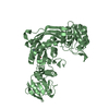
- Structure visualization
| Structure viewer | Molecule: MolmilJmol/JSmol |
|---|
- Downloads & links
-Download
| PDBx/mmCIF format | 7b6q.cif.gz | 375.6 KB | Display | PDBx/mmCIF format |
|---|---|---|---|---|
| PDB format | pdb7b6q.ent.gz | 306.4 KB | Display | PDB format |
| PDBx/mmJSON format | 7b6q.json.gz | Tree view | PDBx/mmJSON format | |
| Others |  Other downloads Other downloads |
-Validation report
| Arichive directory | https://data.pdbj.org/pub/pdb/validation_reports/b6/7b6qftp://data.pdbj.org/pub/pdb/validation_reports/b6/7b6q | HTTPS FTP |
|---|
-Related structure data
| Related structure data |  7b53SC  7b60C  7b61C  7b68C 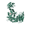 7b6gC 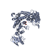 7b6iC 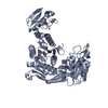 7b6jC  7b6kC 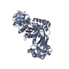 7b6lC 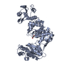 7b6mC  7b6nC  7b6oC  7b6pC  7b9eC 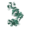 7b9wC S: Starting model for refinement C: citing same article ( |
|---|---|
| Similar structure data |


-Links
 PDBj
PDBj
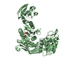
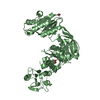
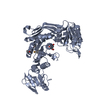
- Assembly
| Deposited unit | 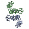
| ||||||||
|---|---|---|---|---|---|---|---|---|---|
| 1 | 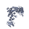
| ||||||||
| 2 | 
| ||||||||
| Unit cell |
|
-Components
| #1: Protein | Mass: 53493.508 Da / Num. of mol.: 2 Source method: isolated from a genetically manipulated source Source: (gene. exp.) Strain: K12 / Gene: murE, b0085, JW0083 / Plasmid: pNIC28-Bsa4 Details (production host): N-terminal His6-tag -Twin-Strep-tag II -TEV-cleavage site Production host: References: UniProt: P22188, UDP-N-acetylmuramoyl-L-alanyl-D-glutamate-2,6-diaminopimelate ligase #2: Chemical | ChemComp-WZD / | 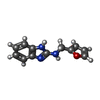  Mass: 213.235 Da / Num. of mol.: 1 / Source method: obtained synthetically / Formula: C12H11N3O / Feature type: SUBJECT OF INVESTIGATION Mass: 213.235 Da / Num. of mol.: 1 / Source method: obtained synthetically / Formula: C12H11N3O / Feature type: SUBJECT OF INVESTIGATION#3: Chemical | ChemComp-IPA / |   Mass: 60.095 Da / Num. of mol.: 1 / Source method: obtained synthetically / Formula: C3H8O Mass: 60.095 Da / Num. of mol.: 1 / Source method: obtained synthetically / Formula: C3H8O#4: Water | ChemComp-HOH / |  Mass: 18.015 Da / Num. of mol.: 287 / Source method: isolated from a natural source / Formula: H2O Mass: 18.015 Da / Num. of mol.: 287 / Source method: isolated from a natural source / Formula: H2OHas ligand of interest | Y | |
|---|
-Experimental details
-Experiment
| Experiment | Method: X-RAY DIFFRACTION / Number of used crystals: 1 |
|---|
- Sample preparation
| Crystal | Density Matthews: 2.24 Å3/Da / Density % sol: 45.19 % |
|---|---|
| Crystal grow | Temperature: 294 K / Method: vapor diffusion, sitting drop / pH: 5.5 / Details: 0.1M citrate pH 5.5 19% PEG4K 14% 2-propanol |
-Data collection
| Diffraction | Mean temperature: 100 K / Serial crystal experiment: N |
|---|---|
| Diffraction source | Source: SYNCHROTRON / Site: Diamond  / Beamline: I04-1 / Wavelength: 0.91587 Å / Beamline: I04-1 / Wavelength: 0.91587 Å |
| Detector | Type: DECTRIS PILATUS3 S 6M / Detector: PIXEL / Date: Jul 13, 2018 |
| Radiation | Protocol: SINGLE WAVELENGTH / Monochromatic (M) / Laue (L): M / Scattering type: x-ray |
| Radiation wavelength | Wavelength: 0.91587 Å / Relative weight: 1 |
| Reflection | Resolution: 1.8→73.55 Å / Num. obs: 82093 / % possible obs: 95 % / Redundancy: 1.8 % / CC1/2: 0.996 / Net I/σ(I): 6.2 |
| Reflection shell | Resolution: 1.8→1.9 Å / Num. unique obs: 12064 / CC1/2: 0.46 |
- Processing
| Software |
| |||||||||||||||||||||||||||||||||||||||||||||||||||||||||||||||||||||||||||||||||||||||||||||||||||||||||
|---|---|---|---|---|---|---|---|---|---|---|---|---|---|---|---|---|---|---|---|---|---|---|---|---|---|---|---|---|---|---|---|---|---|---|---|---|---|---|---|---|---|---|---|---|---|---|---|---|---|---|---|---|---|---|---|---|---|---|---|---|---|---|---|---|---|---|---|---|---|---|---|---|---|---|---|---|---|---|---|---|---|---|---|---|---|---|---|---|---|---|---|---|---|---|---|---|---|---|---|---|---|---|---|---|---|---|
| Refinement | Method to determine structure: MOLECULAR REPLACEMENT Starting model: 7B53 Resolution: 1.82→56.08 Å / Cor.coef. Fo:Fc: 0.956 / Cor.coef. Fo:Fc free: 0.942 / SU B: 8.599 / SU ML: 0.124 / Cross valid method: THROUGHOUT / ESU R: 0.158 / ESU R Free: 0.14 / Stereochemistry target values: MAXIMUM LIKELIHOOD Details: U VALUES : WITH TLS ADDED HYDROGENS HAVE BEEN ADDED IN THE RIDING POSITIONS U VALUES : RESIDUAL ONLY
| |||||||||||||||||||||||||||||||||||||||||||||||||||||||||||||||||||||||||||||||||||||||||||||||||||||||||
| Solvent computation | Ion probe radii: 0.7 Å / Shrinkage radii: 0.7 Å / VDW probe radii: 1.1 Å / Solvent model: MASK | |||||||||||||||||||||||||||||||||||||||||||||||||||||||||||||||||||||||||||||||||||||||||||||||||||||||||
| Displacement parameters | Biso mean: 41.098 Å2
| |||||||||||||||||||||||||||||||||||||||||||||||||||||||||||||||||||||||||||||||||||||||||||||||||||||||||
| Refinement step | Cycle: LAST / Resolution: 1.82→56.08 Å
| |||||||||||||||||||||||||||||||||||||||||||||||||||||||||||||||||||||||||||||||||||||||||||||||||||||||||
| Refine LS restraints |
| |||||||||||||||||||||||||||||||||||||||||||||||||||||||||||||||||||||||||||||||||||||||||||||||||||||||||
| LS refinement shell | Resolution: 1.82→1.867 Å / Total num. of bins used: 20
| |||||||||||||||||||||||||||||||||||||||||||||||||||||||||||||||||||||||||||||||||||||||||||||||||||||||||
| Refinement TLS params. | Method: refined / Refine-ID: X-RAY DIFFRACTION
| |||||||||||||||||||||||||||||||||||||||||||||||||||||||||||||||||||||||||||||||||||||||||||||||||||||||||
| Refinement TLS group |
|
