ムービー
ムービー コントローラー


+ データを開く

- 基本情報
基本情報
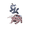
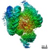
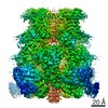
| 登録情報 | データベース: PDB / ID: 7b2l | |||||||||
|---|---|---|---|---|---|---|---|---|---|---|
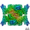
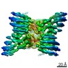
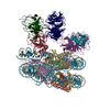
| タイトル | Structure of the endocytic adaptor complex AENTH | |||||||||
 要素 要素 |
| |||||||||
 キーワード キーワード | CELL ADHESION / Membrane protein / clathrin-mediated endocytosis / adapter protein / Sla2 / Epsin-1 / ENTH / ANTH | |||||||||
| 機能・相同性 |  機能・相同性情報 機能・相同性情報Cargo recognition for clathrin-mediated endocytosis / actin cortical patch assembly / clathrin vesicle coat / clathrin light chain binding / incipient cellular bud site / negative regulation of Arp2/3 complex-mediated actin nucleation / cellular bud tip / actin cortical patch / clathrin coat assembly / clathrin adaptor activity ...Cargo recognition for clathrin-mediated endocytosis / actin cortical patch assembly / clathrin vesicle coat / clathrin light chain binding / incipient cellular bud site / negative regulation of Arp2/3 complex-mediated actin nucleation / cellular bud tip / actin cortical patch / clathrin coat assembly / clathrin adaptor activity / cellular bud neck / mating projection tip / phosphatidylinositol-3,4-bisphosphate binding / phosphatidylinositol-3,5-bisphosphate binding / clathrin-coated vesicle / K63-linked polyubiquitin modification-dependent protein binding / clathrin binding / cortical actin cytoskeleton / actin filament organization / ubiquitin binding / phospholipid binding / endocytosis / actin filament binding / early endosome / endosome / plasma membrane / cytoplasm 類似検索 - 分子機能 | |||||||||
| 生物種 |  | |||||||||
| 手法 | 電子顕微鏡法 / 単粒子再構成法 / クライオ電子顕微鏡法 / 解像度: 3.9 Å | |||||||||
 データ登録者 データ登録者 | Klebl, D.P. / Lizarrondo, J. / Sobott, F. / Garcia-Alai, M. / Muench, S.P. | |||||||||
| 資金援助 |  ドイツ, ドイツ,  英国, 2件 英国, 2件
| |||||||||
 引用 引用 | ジャーナル: Nat Commun / 年: 2021 タイトル: Structure of the endocytic adaptor complex reveals the basis for efficient membrane anchoring during clathrin-mediated endocytosis. 著者: Javier Lizarrondo / David P Klebl / Stephan Niebling / Marc Abella / Martin A Schroer / Haydyn D T Mertens / Katharina Veith / Roland Thuenauer / Dmitri I Svergun / Michal Skruzny / Frank ...著者: Javier Lizarrondo / David P Klebl / Stephan Niebling / Marc Abella / Martin A Schroer / Haydyn D T Mertens / Katharina Veith / Roland Thuenauer / Dmitri I Svergun / Michal Skruzny / Frank Sobott / Stephen P Muench / Maria M Garcia-Alai /  要旨: During clathrin-mediated endocytosis, a complex and dynamic network of protein-membrane interactions cooperate to achieve membrane invagination. Throughout this process in yeast, endocytic coat ...During clathrin-mediated endocytosis, a complex and dynamic network of protein-membrane interactions cooperate to achieve membrane invagination. Throughout this process in yeast, endocytic coat adaptors, Sla2 and Ent1, must remain attached to the plasma membrane to transmit force from the actin cytoskeleton required for successful membrane invagination. Here, we present a cryo-EM structure of a 16-mer complex of the ANTH and ENTH membrane-binding domains from Sla2 and Ent1 bound to PIP that constitutes the anchor to the plasma membrane. Detailed in vitro and in vivo mutagenesis of the complex interfaces delineate the key interactions for complex formation and deficient cell growth phenotypes demonstrate its biological relevance. A hetero-tetrameric unit binds PIP molecules at the ANTH-ENTH interfaces and can form larger assemblies to contribute to membrane remodeling. Finally, a time-resolved small-angle X-ray scattering study of the interaction of these adaptor domains in vitro suggests that ANTH and ENTH domains have evolved to achieve a fast subsecond timescale assembly in the presence of PIP and do not require further proteins to form a stable complex. Together, these findings provide a molecular understanding of an essential piece in the molecular puzzle of clathrin-coated endocytic sites. | |||||||||
| 履歴 |
|
- 構造の表示
構造の表示
| ムービー |
ムービービューア |
|---|---|
| 構造ビューア | 分子: MolmilJmol/JSmol |
- ダウンロードとリンク
-ダウンロード
| PDBx/mmCIF形式 | 7b2l.cif.gz | 567.8 KB | 表示 | PDBx/mmCIF形式 |
|---|---|---|---|---|
| PDB形式 | pdb7b2l.ent.gz | 473 KB | 表示 | PDB形式 |
| PDBx/mmJSON形式 | 7b2l.json.gz | ツリー表示 | PDBx/mmJSON形式 | |
| その他 |  その他のダウンロード その他のダウンロード |
-検証レポート
| 文書・要旨 | 7b2l_validation.pdf.gz | 2.4 MB | 表示 | wwPDB検証レポート |
|---|---|---|---|---|
| 文書・詳細版 | 7b2l_full_validation.pdf.gz | 2.4 MB | 表示 | |
| XML形式データ | 7b2l_validation.xml.gz | 92.2 KB | 表示 | |
| CIF形式データ | 7b2l_validation.cif.gz | 139 KB | 表示 | |
| アーカイブディレクトリ | https://data.pdbj.org/pub/pdb/validation_reports/b2/7b2lftp://data.pdbj.org/pub/pdb/validation_reports/b2/7b2l | HTTPS FTP |
-関連構造データ

-リンク
 PDBj
PDBj


- 集合体
集合体
| 登録構造単位 | 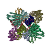
|
|---|---|
| 1 |
|
-要素
| #1: タンパク質 | 分子量: 18880.510 Da / 分子数: 8 / 由来タイプ: 組換発現 / 詳細: ENTH domain of epsin Ent1 由来: (組換発現) 遺伝子: ENT1, YDL161W / 発現宿主:  #2: タンパク質 | 分子量: 33257.004 Da / 分子数: 8 / 由来タイプ: 組換発現 / 詳細: ANTH domain of Sla2 由来: (組換発現) 遺伝子: SLA2, END4, MOP2, UFG1, YNL243W, N1102 / 発現宿主: #3: 化合物 | ChemComp-PIO / [(   分子量: 746.566 Da / 分子数: 20 / 由来タイプ: 合成 / 式: C25H49O19P3 / タイプ: SUBJECT OF INVESTIGATION 分子量: 746.566 Da / 分子数: 20 / 由来タイプ: 合成 / 式: C25H49O19P3 / タイプ: SUBJECT OF INVESTIGATION研究の焦点であるリガンドがあるか | Y | |
|---|
-実験情報
-実験
| 実験 | 手法: 電子顕微鏡法 |
|---|---|
| EM実験 | 試料の集合状態: PARTICLE / 3次元再構成法: 単粒子再構成法 |
- 試料調製
試料調製
| 構成要素 | 名称: 16 mer of 8 Sla2 ANTH and 8 Epsin-1 ENTH domains in complex with PIP2 タイプ: COMPLEX / Entity ID: #1-#2 / 由来: RECOMBINANT |
|---|---|
| 由来(天然) | 生物種: |
| 由来(組換発現) | 生物種: |
| 緩衝液 | pH: 8 |
| 試料 | 濃度: 0.5 mg/ml / 包埋: NO / シャドウイング: NO / 染色: NO / 凍結: YES / 詳細: 20 mM Tris, 250 mM NaCl and 1 mM DTT |
| 急速凍結 | 装置: FEI VITROBOT MARK IV / 凍結剤: ETHANE / 湿度: 90 % / 凍結前の試料温度: 278 K |
- 電子顕微鏡撮影
| 実験機器 |  モデル: Titan Krios / 画像提供: FEI Company |
|---|---|
| 顕微鏡 | モデル: FEI TITAN KRIOS |
| 電子銃 | 電子線源:  FIELD EMISSION GUN / 加速電圧: 300 kV / 照射モード: FLOOD BEAM FIELD EMISSION GUN / 加速電圧: 300 kV / 照射モード: FLOOD BEAM |
| 電子レンズ | モード: BRIGHT FIELD |
| 撮影 | 電子線照射量: 75.2 e/Å2 / 検出モード: INTEGRATING フィルム・検出器のモデル: FEI FALCON III (4k x 4k) |
- 解析
解析
| EMソフトウェア |
| |||||||||||||||||||||||||||
|---|---|---|---|---|---|---|---|---|---|---|---|---|---|---|---|---|---|---|---|---|---|---|---|---|---|---|---|---|
| CTF補正 | タイプ: PHASE FLIPPING AND AMPLITUDE CORRECTION | |||||||||||||||||||||||||||
| 粒子像の選択 | 選択した粒子像数: 195536 | |||||||||||||||||||||||||||
| 3次元再構成 | 解像度: 3.9 Å / 解像度の算出法: FSC 0.143 CUT-OFF / 粒子像の数: 79414 / 対称性のタイプ: POINT | |||||||||||||||||||||||||||
| 原子モデル構築 |
|