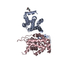
Movie
Movie Controller


+ Open data

- Basic information
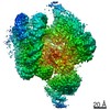
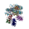
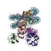
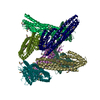
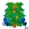
| Entry | Database: PDB / ID: 7b2l | |||||||||
|---|---|---|---|---|---|---|---|---|---|---|
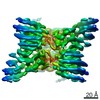
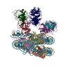
| Title | Structure of the endocytic adaptor complex AENTH | |||||||||
 Components Components |
| |||||||||
 Keywords Keywords | CELL ADHESION / Membrane protein / clathrin-mediated endocytosis / adapter protein / Sla2 / Epsin-1 / ENTH / ANTH | |||||||||
| Function / homology |  Function and homology information Function and homology informationCargo recognition for clathrin-mediated endocytosis / actin cortical patch assembly / clathrin vesicle coat / clathrin light chain binding / incipient cellular bud site / actin cortical patch / cellular bud tip / negative regulation of Arp2/3 complex-mediated actin nucleation / clathrin coat assembly / clathrin-cargo adaptor activity ...Cargo recognition for clathrin-mediated endocytosis / actin cortical patch assembly / clathrin vesicle coat / clathrin light chain binding / incipient cellular bud site / actin cortical patch / cellular bud tip / negative regulation of Arp2/3 complex-mediated actin nucleation / clathrin coat assembly / clathrin-cargo adaptor activity / cellular bud neck / mating projection tip / phosphatidylinositol-3,4-bisphosphate binding / phosphatidylinositol-3,5-bisphosphate binding / clathrin-coated vesicle / K63-linked polyubiquitin modification-dependent protein binding / cortical actin cytoskeleton / clathrin binding / actin filament organization / ubiquitin binding / phospholipid binding / endocytosis / actin filament binding / early endosome / endosome / plasma membrane / cytoplasm Similarity search - Function | |||||||||
| Biological species |  | |||||||||
| Method | ELECTRON MICROSCOPY / single particle reconstruction / cryo EM / Resolution: 3.9 Å | |||||||||
 Authors Authors | Klebl, D.P. / Lizarrondo, J. / Sobott, F. / Garcia-Alai, M. / Muench, S.P. | |||||||||
| Funding support |  Germany, Germany,  United Kingdom, 2items United Kingdom, 2items
| |||||||||
 Citation Citation | Journal: Nat Commun / Year: 2021 Title: Structure of the endocytic adaptor complex reveals the basis for efficient membrane anchoring during clathrin-mediated endocytosis. Authors: Javier Lizarrondo / David P Klebl / Stephan Niebling / Marc Abella / Martin A Schroer / Haydyn D T Mertens / Katharina Veith / Roland Thuenauer / Dmitri I Svergun / Michal Skruzny / Frank ...Authors: Javier Lizarrondo / David P Klebl / Stephan Niebling / Marc Abella / Martin A Schroer / Haydyn D T Mertens / Katharina Veith / Roland Thuenauer / Dmitri I Svergun / Michal Skruzny / Frank Sobott / Stephen P Muench / Maria M Garcia-Alai /  Abstract: During clathrin-mediated endocytosis, a complex and dynamic network of protein-membrane interactions cooperate to achieve membrane invagination. Throughout this process in yeast, endocytic coat ...During clathrin-mediated endocytosis, a complex and dynamic network of protein-membrane interactions cooperate to achieve membrane invagination. Throughout this process in yeast, endocytic coat adaptors, Sla2 and Ent1, must remain attached to the plasma membrane to transmit force from the actin cytoskeleton required for successful membrane invagination. Here, we present a cryo-EM structure of a 16-mer complex of the ANTH and ENTH membrane-binding domains from Sla2 and Ent1 bound to PIP that constitutes the anchor to the plasma membrane. Detailed in vitro and in vivo mutagenesis of the complex interfaces delineate the key interactions for complex formation and deficient cell growth phenotypes demonstrate its biological relevance. A hetero-tetrameric unit binds PIP molecules at the ANTH-ENTH interfaces and can form larger assemblies to contribute to membrane remodeling. Finally, a time-resolved small-angle X-ray scattering study of the interaction of these adaptor domains in vitro suggests that ANTH and ENTH domains have evolved to achieve a fast subsecond timescale assembly in the presence of PIP and do not require further proteins to form a stable complex. Together, these findings provide a molecular understanding of an essential piece in the molecular puzzle of clathrin-coated endocytic sites. | |||||||||
| History |
|
- Structure visualization
| Movie |
Movie viewer |
|---|---|
| Structure viewer | Molecule: MolmilJmol/JSmol |
- Downloads & links
-Download
| PDBx/mmCIF format | 7b2l.cif.gz | 567.8 KB | Display | PDBx/mmCIF format |
|---|---|---|---|---|
| PDB format | pdb7b2l.ent.gz | 473 KB | Display | PDB format |
| PDBx/mmJSON format | 7b2l.json.gz | Tree view | PDBx/mmJSON format | |
| Others |  Other downloads Other downloads |
-Validation report
| Arichive directory | https://data.pdbj.org/pub/pdb/validation_reports/b2/7b2lftp://data.pdbj.org/pub/pdb/validation_reports/b2/7b2l | HTTPS FTP |
|---|
-Related structure data
| Related structure data | 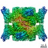 11987MC C: citing same article ( M: map data used to model this data |
|---|---|
| Similar structure data |

-Links
 PDBj
PDBj


- Assembly
| Deposited unit | 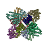
|
|---|---|
| 1 |
|
-Components
| #1: Protein | Mass: 18880.510 Da / Num. of mol.: 8 Source method: isolated from a genetically manipulated source Details: ENTH domain of epsin Ent1 / Source: (gene. exp.)  #2: Protein | Mass: 33257.004 Da / Num. of mol.: 8 Source method: isolated from a genetically manipulated source Details: ANTH domain of Sla2 / Source: (gene. exp.) #3: Chemical | ChemComp-PIO / [(   Mass: 746.566 Da / Num. of mol.: 20 / Source method: obtained synthetically / Formula: C25H49O19P3 / Feature type: SUBJECT OF INVESTIGATION Mass: 746.566 Da / Num. of mol.: 20 / Source method: obtained synthetically / Formula: C25H49O19P3 / Feature type: SUBJECT OF INVESTIGATIONHas ligand of interest | Y | |
|---|
-Experimental details
-Experiment
| Experiment | Method: ELECTRON MICROSCOPY |
|---|---|
| EM experiment | Aggregation state: PARTICLE / 3D reconstruction method: single particle reconstruction |
- Sample preparation
| Component | Name: 16 mer of 8 Sla2 ANTH and 8 Epsin-1 ENTH domains in complex with PIP2 Type: COMPLEX / Entity ID: #1-#2 / Source: RECOMBINANT |
|---|---|
| Source (natural) | Organism: |
| Source (recombinant) | Organism: |
| Buffer solution | pH: 8 |
| Specimen | Conc.: 0.5 mg/ml / Embedding applied: NO / Shadowing applied: NO / Staining applied: NO / Vitrification applied: YES / Details: 20 mM Tris, 250 mM NaCl and 1 mM DTT |
| Vitrification | Instrument: FEI VITROBOT MARK IV / Cryogen name: ETHANE / Humidity: 90 % / Chamber temperature: 278 K |
- Electron microscopy imaging
| Experimental equipment |  Model: Titan Krios / Image courtesy: FEI Company |
|---|---|
| Microscopy | Model: FEI TITAN KRIOS |
| Electron gun | Electron source:  FIELD EMISSION GUN / Accelerating voltage: 300 kV / Illumination mode: FLOOD BEAM FIELD EMISSION GUN / Accelerating voltage: 300 kV / Illumination mode: FLOOD BEAM |
| Electron lens | Mode: BRIGHT FIELD |
| Image recording | Electron dose: 75.2 e/Å2 / Detector mode: INTEGRATING / Film or detector model: FEI FALCON III (4k x 4k) |
- Processing
| EM software |
| |||||||||||||||||||||||||||
|---|---|---|---|---|---|---|---|---|---|---|---|---|---|---|---|---|---|---|---|---|---|---|---|---|---|---|---|---|
| CTF correction | Type: PHASE FLIPPING AND AMPLITUDE CORRECTION | |||||||||||||||||||||||||||
| Particle selection | Num. of particles selected: 195536 | |||||||||||||||||||||||||||
| 3D reconstruction | Resolution: 3.9 Å / Resolution method: FSC 0.143 CUT-OFF / Num. of particles: 79414 / Symmetry type: POINT | |||||||||||||||||||||||||||
| Atomic model building |
|