 Movie
Movie Controller

[English] 日本語
 Yorodumi
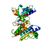
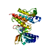
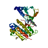
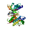
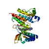
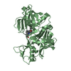
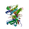
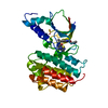
- PDB-7aw2: MerTK kinase domain with type 1.5 inhibitor from a DNA-encoded library -
+ Open data

- Basic information
| Entry | Database: PDB / ID: 7aw2 | ||||||
|---|---|---|---|---|---|---|---|
| Title | MerTK kinase domain with type 1.5 inhibitor from a DNA-encoded library | ||||||
 Components Components | Tyrosine-protein kinase Mer | ||||||
 Keywords Keywords | SIGNALING PROTEIN / tyrosine kinase / inhibitor / type1.5 kinase inhibitor / structure-based drug design / DNA-encoded library / oncology | ||||||
| Function / homology |  Function and homology information Function and homology informationnegative regulation of leukocyte apoptotic process / negative regulation of lymphocyte activation / neutrophil clearance / natural killer cell differentiation / secretion by cell / negative regulation of cytokine production / vagina development / photoreceptor outer segment / establishment of localization in cell / phagocytosis ...negative regulation of leukocyte apoptotic process / negative regulation of lymphocyte activation / neutrophil clearance / natural killer cell differentiation / secretion by cell / negative regulation of cytokine production / vagina development / photoreceptor outer segment / establishment of localization in cell / phagocytosis / substrate adhesion-dependent cell spreading / transmembrane receptor protein tyrosine kinase activity / positive regulation of phagocytosis / cell surface receptor protein tyrosine kinase signaling pathway / Cell surface interactions at the vascular wall / receptor protein-tyrosine kinase / platelet activation / cell-cell signaling / nervous system development / cell migration / retina development in camera-type eye / spermatogenesis / protein phosphorylation / positive regulation of phosphatidylinositol 3-kinase/protein kinase B signal transduction / cell surface receptor signaling pathway / signaling receptor complex / : / ATP binding / plasma membrane / cytoplasm Similarity search - Function | ||||||
| Biological species |  Homo sapiens (human) Homo sapiens (human) | ||||||
| Method |  X-RAY DIFFRACTION / SYNCHROTRON / MOLECULAR REPLACEMENT / molecular replacement / Resolution: 2.1 Å X-RAY DIFFRACTION / SYNCHROTRON / MOLECULAR REPLACEMENT / molecular replacement / Resolution: 2.1 Å | ||||||
 Authors Authors | Schimpl, M. / Nissink, J.W.M. / Blackett, C. / Goldberg, K. / Hennessy, E.J. / Hardaker, E. / McCoull, W. / McMurray, L. / Collingwood, O. / Overman, R. ...Schimpl, M. / Nissink, J.W.M. / Blackett, C. / Goldberg, K. / Hennessy, E.J. / Hardaker, E. / McCoull, W. / McMurray, L. / Collingwood, O. / Overman, R. / Pflug, A. / Preston, M. / Rawlins, P. / Rivers, E. / Smith, P. / Underwood, E. / Truman, C. / Warwicker, J. / Winter, J. / Woodcock, S. | ||||||
 Citation Citation | Journal: J.Med.Chem. / Year: 2021 Title: Generating Selective Leads for Mer Kinase Inhibitors-Example of a Comprehensive Lead-Generation Strategy. Authors: Nissink, J.W.M. / Bazzaz, S. / Blackett, C. / Clark, M.A. / Collingwood, O. / Disch, J.S. / Gikunju, D. / Goldberg, K. / Guilinger, J.P. / Hardaker, E. / Hennessy, E.J. / Jetson, R. / Keefe, ...Authors: Nissink, J.W.M. / Bazzaz, S. / Blackett, C. / Clark, M.A. / Collingwood, O. / Disch, J.S. / Gikunju, D. / Goldberg, K. / Guilinger, J.P. / Hardaker, E. / Hennessy, E.J. / Jetson, R. / Keefe, A.D. / McCoull, W. / McMurray, L. / Olszewski, A. / Overman, R. / Pflug, A. / Preston, M. / Rawlins, P.B. / Rivers, E. / Schimpl, M. / Smith, P. / Truman, C. / Underwood, E. / Warwicker, J. / Winter-Holt, J. / Woodcock, S. / Zhang, Y. | ||||||
| History |
|
- Structure visualization
| Structure viewer | Molecule: MolmilJmol/JSmol |
|---|
- Downloads & links
-Download
| PDBx/mmCIF format | 7aw2.cif.gz | 125.7 KB | Display | PDBx/mmCIF format |
|---|---|---|---|---|
| PDB format | pdb7aw2.ent.gz | 95.6 KB | Display | PDB format |
| PDBx/mmJSON format | 7aw2.json.gz | Tree view | PDBx/mmJSON format | |
| Others |  Other downloads Other downloads |
-Validation report
| Arichive directory | https://data.pdbj.org/pub/pdb/validation_reports/aw/7aw2ftp://data.pdbj.org/pub/pdb/validation_reports/aw/7aw2 | HTTPS FTP |
|---|
-Related structure data
| Related structure data |  7avxC  7avyC  7avzC  7aw0C  7aw1C  7aw3C  7aw4C C: citing same article ( |
|---|---|
| Similar structure data |


-Links
 PDBj
PDBj


- Assembly
| Deposited unit | 
| ||||||||
|---|---|---|---|---|---|---|---|---|---|
| 1 |
| ||||||||
| Unit cell |
|
-Components
| #1: Protein | Mass: 34381.754 Da / Num. of mol.: 1 / Fragment: kinase domain (571-864) / Mutation: K591R,K693R,K702R,K856R Source method: isolated from a genetically manipulated source Details: Co-expressed with PTP1b / Source: (gene. exp.) Homo sapiens (human) / Gene: MERTK, MER / Production host:  References: UniProt: Q12866, receptor protein-tyrosine kinase |
|---|---|
| #2: Chemical | ChemComp-S4W /   Mass: 415.875 Da / Num. of mol.: 1 / Source method: obtained synthetically / Formula: C23H18ClN5O / Feature type: SUBJECT OF INVESTIGATION Mass: 415.875 Da / Num. of mol.: 1 / Source method: obtained synthetically / Formula: C23H18ClN5O / Feature type: SUBJECT OF INVESTIGATION |
| #3: Water | ChemComp-HOH /  Mass: 18.015 Da / Num. of mol.: 69 / Source method: isolated from a natural source / Formula: H2O Mass: 18.015 Da / Num. of mol.: 69 / Source method: isolated from a natural source / Formula: H2O |
| Has ligand of interest | Y |
-Experimental details
-Experiment
| Experiment | Method: X-RAY DIFFRACTION / Number of used crystals: 1 |
|---|
- Sample preparation
| Crystal | Density Matthews: 2.26 Å3/Da / Density % sol: 45.56 % |
|---|---|
| Crystal grow | Temperature: 293 K / Method: vapor diffusion / pH: 8.5 Details: 0.5 mM protein was crystallised in 4.3 M sodium chloride, 0.1 M Tris pH 8.5 and soaked for 24 h with 20 mM compound and 20 % DMSO |
-Data collection
| Diffraction | Mean temperature: 100 K / Serial crystal experiment: N | ||||||||||||||||||||||||||||||
|---|---|---|---|---|---|---|---|---|---|---|---|---|---|---|---|---|---|---|---|---|---|---|---|---|---|---|---|---|---|---|---|
| Diffraction source | Source: SYNCHROTRON / Site: SOLEIL  / Beamline: PROXIMA 2 / Wavelength: 0.9801 Å / Beamline: PROXIMA 2 / Wavelength: 0.9801 Å | ||||||||||||||||||||||||||||||
| Detector | Type: DECTRIS EIGER2 X 9M / Detector: PIXEL / Date: Nov 23, 2016 | ||||||||||||||||||||||||||||||
| Radiation | Protocol: SINGLE WAVELENGTH / Monochromatic (M) / Laue (L): M / Scattering type: x-ray | ||||||||||||||||||||||||||||||
| Radiation wavelength | Wavelength: 0.9801 Å / Relative weight: 1 | ||||||||||||||||||||||||||||||
| Reflection | Resolution: 2.1→48.44 Å / Num. obs: 18583 / % possible obs: 99.7 % / Redundancy: 6.7 % / Biso Wilson estimate: 57.22 Å2 / CC1/2: 0.999 / Rmerge(I) obs: 0.059 / Rpim(I) all: 0.025 / Rrim(I) all: 0.064 / Net I/σ(I): 15.8 / Num. measured all: 124104 | ||||||||||||||||||||||||||||||
| Reflection shell | Diffraction-ID: 1
|
-Phasing
| Phasing | Method: molecular replacement |
|---|
- Processing
| Software |
| ||||||||||||||||||||||||||||||||||||||||||||||||||||||||||||||||||||||||||||||||||||||||||||||||||||||||||||
|---|---|---|---|---|---|---|---|---|---|---|---|---|---|---|---|---|---|---|---|---|---|---|---|---|---|---|---|---|---|---|---|---|---|---|---|---|---|---|---|---|---|---|---|---|---|---|---|---|---|---|---|---|---|---|---|---|---|---|---|---|---|---|---|---|---|---|---|---|---|---|---|---|---|---|---|---|---|---|---|---|---|---|---|---|---|---|---|---|---|---|---|---|---|---|---|---|---|---|---|---|---|---|---|---|---|---|---|---|---|
| Refinement | Method to determine structure: MOLECULAR REPLACEMENT Starting model: internal model Resolution: 2.1→20.73 Å / Cor.coef. Fo:Fc: 0.943 / Cor.coef. Fo:Fc free: 0.937 / SU R Cruickshank DPI: 0.206 / Cross valid method: THROUGHOUT / σ(F): 0 / SU R Blow DPI: 0.209 / SU Rfree Blow DPI: 0.171 / SU Rfree Cruickshank DPI: 0.172
| ||||||||||||||||||||||||||||||||||||||||||||||||||||||||||||||||||||||||||||||||||||||||||||||||||||||||||||
| Displacement parameters | Biso max: 139.65 Å2 / Biso mean: 65.54 Å2 / Biso min: 29.82 Å2
| ||||||||||||||||||||||||||||||||||||||||||||||||||||||||||||||||||||||||||||||||||||||||||||||||||||||||||||
| Refine analyze | Luzzati coordinate error obs: 0.31 Å | ||||||||||||||||||||||||||||||||||||||||||||||||||||||||||||||||||||||||||||||||||||||||||||||||||||||||||||
| Refinement step | Cycle: final / Resolution: 2.1→20.73 Å
| ||||||||||||||||||||||||||||||||||||||||||||||||||||||||||||||||||||||||||||||||||||||||||||||||||||||||||||
| Refine LS restraints |
| ||||||||||||||||||||||||||||||||||||||||||||||||||||||||||||||||||||||||||||||||||||||||||||||||||||||||||||
| LS refinement shell | Resolution: 2.1→2.11 Å / Rfactor Rfree error: 0 / Total num. of bins used: 46
| ||||||||||||||||||||||||||||||||||||||||||||||||||||||||||||||||||||||||||||||||||||||||||||||||||||||||||||
| Refinement TLS params. | Method: refined / Origin x: 15.2423 Å / Origin y: 29.0035 Å / Origin z: -7.6245 Å
| ||||||||||||||||||||||||||||||||||||||||||||||||||||||||||||||||||||||||||||||||||||||||||||||||||||||||||||
| Refinement TLS group | Selection details: { A|* } |
