 Movie
Movie Controller


+ Open data

- Basic information
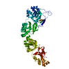
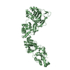
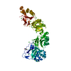
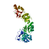
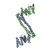
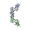
| Entry | Database: PDB / ID: 7asg | ||||||
|---|---|---|---|---|---|---|---|
| Title | TGFBIp mutant R555W | ||||||
 Components Components | Transforming growth factor-beta-induced protein ig-h3 | ||||||
 Keywords Keywords | PROTEIN FIBRIL / granular corneal dystrophy / lattice corneal dystrophy / protein aggregation / extracellular matrix protein | ||||||
| Function / homology |  Function and homology information Function and homology informationnegative regulation of cell adhesion / extracellular matrix binding / extracellular matrix structural constituent / basement membrane / chondrocyte differentiation / collagen binding / extracellular matrix organization / visual perception / cell adhesion molecule binding / trans-Golgi network ...negative regulation of cell adhesion / extracellular matrix binding / extracellular matrix structural constituent / basement membrane / chondrocyte differentiation / collagen binding / extracellular matrix organization / visual perception / cell adhesion molecule binding / trans-Golgi network / integrin binding / extracellular matrix / angiogenesis / cell adhesion / cell population proliferation / Amyloid fiber formation / : / extracellular exosome / extracellular region / identical protein binding / plasma membrane Similarity search - Function | ||||||
| Biological species |  Homo sapiens (human) Homo sapiens (human) | ||||||
| Method |  X-RAY DIFFRACTION / SYNCHROTRON / MOLECULAR REPLACEMENT / Resolution: 2 Å X-RAY DIFFRACTION / SYNCHROTRON / MOLECULAR REPLACEMENT / Resolution: 2 Å | ||||||
 Authors Authors | Nielsen, N.S. / Gadeberg, T.A.F. / Andersen, G.R. | ||||||
 Citation Citation | Journal: J.Biol.Chem. / Year: 2021 Title: Mutation-induced dimerization of transforming growth factor-beta-induced protein may drive protein aggregation in granular corneal dystrophy. Authors: Nielsen, N.S. / Gadeberg, T.A.F. / Poulsen, E.T. / Harwood, S.L. / Weberskov, C.E. / Pedersen, J.S. / Andersen, G.R. / Enghild, J.J. | ||||||
| History |
|
- Structure visualization
| Structure viewer | Molecule: MolmilJmol/JSmol |
|---|
- Downloads & links
-Download
| PDBx/mmCIF format | 7asg.cif.gz | 416.3 KB | Display | PDBx/mmCIF format |
|---|---|---|---|---|
| PDB format | pdb7asg.ent.gz | 287.2 KB | Display | PDB format |
| PDBx/mmJSON format | 7asg.json.gz | Tree view | PDBx/mmJSON format | |
| Others |  Other downloads Other downloads |
-Validation report
| Arichive directory | https://data.pdbj.org/pub/pdb/validation_reports/as/7asgftp://data.pdbj.org/pub/pdb/validation_reports/as/7asg | HTTPS FTP |
|---|
-Related structure data
| Related structure data |  7as7C  7ascC  5nv6S S: Starting model for refinement C: citing same article ( |
|---|---|
| Similar structure data |


-Links
 PDBj
PDBj- Assembly
| Deposited unit | 
| ||||||||||||
|---|---|---|---|---|---|---|---|---|---|---|---|---|---|
| 1 |
| ||||||||||||
| Unit cell |
| ||||||||||||
| Components on special symmetry positions |
|
-Components
| #1: Protein | Mass: 65132.379 Da / Num. of mol.: 1 / Mutation: R555W Source method: isolated from a genetically manipulated source Source: (gene. exp.) Homo sapiens (human) / Gene: TGFBI, BIGH3 / Cell line (production host): HEK293 / Production host: Homo sapiens (human) / References: UniProt: Q15582 |
|---|---|
| #2: Water | ChemComp-HOH /  Mass: 18.015 Da / Num. of mol.: 256 / Source method: isolated from a natural source / Formula: H2O Mass: 18.015 Da / Num. of mol.: 256 / Source method: isolated from a natural source / Formula: H2O |
| Has protein modification | Y |
-Experimental details
-Experiment
| Experiment | Method: X-RAY DIFFRACTION / Number of used crystals: 1 |
|---|
- Sample preparation
| Crystal | Density Matthews: 3.07 Å3/Da / Density % sol: 59.89 % |
|---|---|
| Crystal grow | Temperature: 292 K / Method: vapor diffusion / pH: 7 Details: 0.5 M Succinic acid pH 7.0, 0.1 M BIS-TRIS propane pH 7.0 |
-Data collection
| Diffraction | Mean temperature: 100 K / Serial crystal experiment: N |
|---|---|
| Diffraction source | Source: SYNCHROTRON / Site: PETRA III, EMBL c/o DESY  / Beamline: P14 (MX2) / Wavelength: 0.9763 Å / Beamline: P14 (MX2) / Wavelength: 0.9763 Å |
| Detector | Type: DECTRIS EIGER X 16M / Detector: PIXEL / Date: Jun 2, 2018 |
| Radiation | Protocol: SINGLE WAVELENGTH / Monochromatic (M) / Laue (L): M / Scattering type: x-ray |
| Radiation wavelength | Wavelength: 0.9763 Å / Relative weight: 1 |
| Reflection | Resolution: 2→49.24 Å / Num. obs: 54442 / % possible obs: 99.82 % / Redundancy: 10.4 % / Biso Wilson estimate: 40.78 Å2 / CC1/2: 0.998 / CC star: 0.999 / Rmerge(I) obs: 0.1883 / Rrim(I) all: 0.198 / Net I/σ(I): 8.38 |
| Reflection shell | Resolution: 2→2.071 Å / Redundancy: 10.5 % / Num. unique obs: 5356 / CC1/2: 0.53 / CC star: 0.833 / % possible all: 99.6 |
- Processing
| Software |
| |||||||||||||||||||||||||||||||||||||||||||||||||||||||||||||||||||||||||||||||||||||||||||||||||||||||||||||||||||||||||||||||||||||||||||||||||||||||||||||||||||||||||||||||
|---|---|---|---|---|---|---|---|---|---|---|---|---|---|---|---|---|---|---|---|---|---|---|---|---|---|---|---|---|---|---|---|---|---|---|---|---|---|---|---|---|---|---|---|---|---|---|---|---|---|---|---|---|---|---|---|---|---|---|---|---|---|---|---|---|---|---|---|---|---|---|---|---|---|---|---|---|---|---|---|---|---|---|---|---|---|---|---|---|---|---|---|---|---|---|---|---|---|---|---|---|---|---|---|---|---|---|---|---|---|---|---|---|---|---|---|---|---|---|---|---|---|---|---|---|---|---|---|---|---|---|---|---|---|---|---|---|---|---|---|---|---|---|---|---|---|---|---|---|---|---|---|---|---|---|---|---|---|---|---|---|---|---|---|---|---|---|---|---|---|---|---|---|---|---|---|---|
| Refinement | Method to determine structure: MOLECULAR REPLACEMENT Starting model: 5nv6 Resolution: 2→49.24 Å / SU ML: 0.3219 / Cross valid method: FREE R-VALUE / σ(F): 1.34 / Phase error: 29.2167
| |||||||||||||||||||||||||||||||||||||||||||||||||||||||||||||||||||||||||||||||||||||||||||||||||||||||||||||||||||||||||||||||||||||||||||||||||||||||||||||||||||||||||||||||
| Solvent computation | Shrinkage radii: 0.9 Å / VDW probe radii: 1.11 Å / Solvent model: FLAT BULK SOLVENT MODEL | |||||||||||||||||||||||||||||||||||||||||||||||||||||||||||||||||||||||||||||||||||||||||||||||||||||||||||||||||||||||||||||||||||||||||||||||||||||||||||||||||||||||||||||||
| Displacement parameters | Biso mean: 55.76 Å2 | |||||||||||||||||||||||||||||||||||||||||||||||||||||||||||||||||||||||||||||||||||||||||||||||||||||||||||||||||||||||||||||||||||||||||||||||||||||||||||||||||||||||||||||||
| Refinement step | Cycle: LAST / Resolution: 2→49.24 Å
| |||||||||||||||||||||||||||||||||||||||||||||||||||||||||||||||||||||||||||||||||||||||||||||||||||||||||||||||||||||||||||||||||||||||||||||||||||||||||||||||||||||||||||||||
| Refine LS restraints |
| |||||||||||||||||||||||||||||||||||||||||||||||||||||||||||||||||||||||||||||||||||||||||||||||||||||||||||||||||||||||||||||||||||||||||||||||||||||||||||||||||||||||||||||||
| LS refinement shell |
| |||||||||||||||||||||||||||||||||||||||||||||||||||||||||||||||||||||||||||||||||||||||||||||||||||||||||||||||||||||||||||||||||||||||||||||||||||||||||||||||||||||||||||||||
| Refinement TLS params. | Method: refined / Refine-ID: X-RAY DIFFRACTION
| |||||||||||||||||||||||||||||||||||||||||||||||||||||||||||||||||||||||||||||||||||||||||||||||||||||||||||||||||||||||||||||||||||||||||||||||||||||||||||||||||||||||||||||||
| Refinement TLS group |
|
