 Movie
Movie Controller


+ Open data

- Basic information
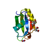
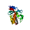
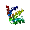
| Entry | Database: PDB / ID: 7arr | ||||||
|---|---|---|---|---|---|---|---|
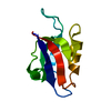
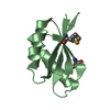
| Title | The de novo designed hybrid alpha/beta-miniprotein | ||||||
 Components Components | alpha/beta-peptide | ||||||
 Keywords Keywords | DE NOVO PROTEIN / alpha/beta-peptide / foldamer | ||||||
| Function / homology | trifluoroacetic acid Function and homology information Function and homology information | ||||||
| Biological species | synthetic construct (others) | ||||||
| Method |  X-RAY DIFFRACTION / SYNCHROTRON / MOLECULAR REPLACEMENT / Resolution: 1.1 Å X-RAY DIFFRACTION / SYNCHROTRON / MOLECULAR REPLACEMENT / Resolution: 1.1 Å | ||||||
 Authors Authors | Bejger, M. / Fortuna, P. / Drewniak-Switalska, M. / Rypniewski, W. / Berlicki, L. | ||||||
| Funding support |  Poland, 1items Poland, 1items
| ||||||
 Citation Citation | Journal: Chem.Commun.(Camb.) / Year: 2021 Title: A computationally designed beta-amino acid-containing miniprotein. Authors: Bejger, M. / Fortuna, P. / Drewniak-Switalska, M. / Plewka, J. / Rypniewski, W. / Berlicki, L. | ||||||
| History |
|
- Structure visualization
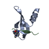
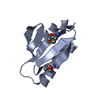
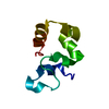
| Structure viewer | Molecule: MolmilJmol/JSmol |
|---|
- Downloads & links
-Download
| PDBx/mmCIF format | 7arr.cif.gz | 89.2 KB | Display | PDBx/mmCIF format |
|---|---|---|---|---|
| PDB format | pdb7arr.ent.gz | 70.5 KB | Display | PDB format |
| PDBx/mmJSON format | 7arr.json.gz | Tree view | PDBx/mmJSON format | |
| Others |  Other downloads Other downloads |
-Validation report
| Arichive directory | https://data.pdbj.org/pub/pdb/validation_reports/ar/7arrftp://data.pdbj.org/pub/pdb/validation_reports/ar/7arr | HTTPS FTP |
|---|
-Related structure data
| Related structure data | 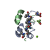 7arsC C: citing same article ( |
|---|---|
| Similar structure data | |
| Experimental dataset #1 | Data reference: 10.18150/EM87YL / Data set type: diffraction image data / Details: MX-RDR, V1 |

-Links
 PDBj
PDBj
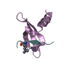
- Assembly
| Deposited unit | 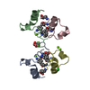
| ||||||||
|---|---|---|---|---|---|---|---|---|---|
| 1 | 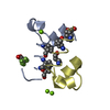
| ||||||||
| 2 | 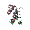
| ||||||||
| Unit cell |
|
-Components
| #1: Protein/peptide | Mass: 4081.618 Da / Num. of mol.: 4 / Source method: obtained synthetically / Source: (synth.) synthetic construct (others) #2: Chemical | ChemComp-TFA / | 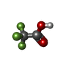  Mass: 114.023 Da / Num. of mol.: 1 / Source method: obtained synthetically / Formula: C2HF3O2 Mass: 114.023 Da / Num. of mol.: 1 / Source method: obtained synthetically / Formula: C2HF3O2#3: Chemical |   Mass: 24.305 Da / Num. of mol.: 3 / Source method: isolated from a natural source / Formula: Mg Mass: 24.305 Da / Num. of mol.: 3 / Source method: isolated from a natural source / Formula: Mg#4: Chemical | ChemComp-CL / |   Mass: 35.453 Da / Num. of mol.: 1 / Source method: obtained synthetically / Formula: Cl Mass: 35.453 Da / Num. of mol.: 1 / Source method: obtained synthetically / Formula: Cl#5: Water | ChemComp-HOH / |  Mass: 18.015 Da / Num. of mol.: 298 / Source method: isolated from a natural source / Formula: H2O Mass: 18.015 Da / Num. of mol.: 298 / Source method: isolated from a natural source / Formula: H2OHas ligand of interest | N | |
|---|
-Experimental details
-Experiment
| Experiment | Method: X-RAY DIFFRACTION / Number of used crystals: 1 |
|---|
- Sample preparation
| Crystal | Density Matthews: 2.28 Å3/Da / Density % sol: 46.01 % |
|---|---|
| Crystal grow | Temperature: 291 K / Method: vapor diffusion, hanging drop / pH: 7.5 Details: 0.2 M magnesium chloride hexahydrate 0.1 M HEPES pH 7.5 25 % w/v PEG 3350 |
-Data collection
| Diffraction | Mean temperature: 100 K / Serial crystal experiment: N |
|---|---|
| Diffraction source | Source: SYNCHROTRON / Site: PETRA III, EMBL c/o DESY  / Beamline: P13 (MX1) / Wavelength: 0.97626 Å / Beamline: P13 (MX1) / Wavelength: 0.97626 Å |
| Detector | Type: DECTRIS PILATUS 6M / Detector: PIXEL / Date: Dec 1, 2018 |
| Radiation | Protocol: SINGLE WAVELENGTH / Monochromatic (M) / Laue (L): M / Scattering type: x-ray |
| Radiation wavelength | Wavelength: 0.97626 Å / Relative weight: 1 |
| Reflection | Resolution: 1.1→50 Å / Num. obs: 49390 / % possible obs: 87.3 % / Redundancy: 3.5 % / Biso Wilson estimate: 15.1 Å2 / CC1/2: 0.99 / Rmerge(I) obs: 0.027 / Net I/σ(I): 24.5 |
| Reflection shell | Resolution: 1.1→1.17 Å / Redundancy: 3.5 % / Rmerge(I) obs: 0.14 / Mean I/σ(I) obs: 7.34 / Num. unique obs: 7297 / CC1/2: 0.98 / % possible all: 80.1 |
- Processing
| Software |
| |||||||||||||||||||||||||||||||||||||||||||||||||||||||||||||||||
|---|---|---|---|---|---|---|---|---|---|---|---|---|---|---|---|---|---|---|---|---|---|---|---|---|---|---|---|---|---|---|---|---|---|---|---|---|---|---|---|---|---|---|---|---|---|---|---|---|---|---|---|---|---|---|---|---|---|---|---|---|---|---|---|---|---|---|
| Refinement | Method to determine structure: MOLECULAR REPLACEMENT Starting model: not deposited Resolution: 1.1→29.02 Å / Cor.coef. Fo:Fc: 0.982 / Cor.coef. Fo:Fc free: 0.975 / SU B: 1.174 / SU ML: 0.024 / Cross valid method: THROUGHOUT / σ(F): 0 / ESU R: 0.032 / ESU R Free: 0.034 / Stereochemistry target values: MAXIMUM LIKELIHOOD Details: HYDROGENS HAVE BEEN ADDED IN THE RIDING POSITIONS U VALUES : REFINED INDIVIDUALLY
| |||||||||||||||||||||||||||||||||||||||||||||||||||||||||||||||||
| Solvent computation | Ion probe radii: 0.8 Å / Shrinkage radii: 0.8 Å / VDW probe radii: 1.2 Å / Solvent model: MASK | |||||||||||||||||||||||||||||||||||||||||||||||||||||||||||||||||
| Displacement parameters | Biso max: 101.16 Å2 / Biso mean: 14.162 Å2 / Biso min: 6.17 Å2
| |||||||||||||||||||||||||||||||||||||||||||||||||||||||||||||||||
| Refinement step | Cycle: final / Resolution: 1.1→29.02 Å
| |||||||||||||||||||||||||||||||||||||||||||||||||||||||||||||||||
| Refine LS restraints |
| |||||||||||||||||||||||||||||||||||||||||||||||||||||||||||||||||
| LS refinement shell | Resolution: 1.1→1.129 Å / Rfactor Rfree error: 0 / Total num. of bins used: 20
|
