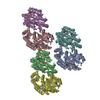
Movie
Movie Controller


+ Open data

- Basic information
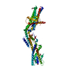
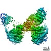
| Entry | Database: PDB / ID: 7anz | ||||||
|---|---|---|---|---|---|---|---|
| Title | Structure of the Candida albicans gamma-Tubulin Small Complex | ||||||
 Components Components |
| ||||||
 Keywords Keywords | CYTOSOLIC PROTEIN / gamma-Tubulin Small Complex / Cytoskeleton / Microtubule nucleation | ||||||
| Function / homology |  Function and homology information Function and homology informationinner plaque of spindle pole body / microtubule nucleation by spindle pole body / outer plaque of spindle pole body / gamma-tubulin small complex / gamma-tubulin complex / microtubule nucleation / spindle pole body / gamma-tubulin binding / positive regulation of cytoplasmic translation / spindle assembly ...inner plaque of spindle pole body / microtubule nucleation by spindle pole body / outer plaque of spindle pole body / gamma-tubulin small complex / gamma-tubulin complex / microtubule nucleation / spindle pole body / gamma-tubulin binding / positive regulation of cytoplasmic translation / spindle assembly / cytoplasmic microtubule organization / mitotic spindle organization / meiotic cell cycle / structural constituent of cytoskeleton / spindle pole / mitotic cell cycle / microtubule / GTP binding Similarity search - Function | ||||||
| Biological species |  Candida albicans (yeast) Candida albicans (yeast) | ||||||
| Method | ELECTRON MICROSCOPY / single particle reconstruction / cryo EM / Resolution: 3.6 Å | ||||||
 Authors Authors | Zupa, E. / Pfeffer, S. | ||||||
| Funding support |  Germany, 1items Germany, 1items
| ||||||
 Citation Citation | Journal: Nat Commun / Year: 2020 Title: The cryo-EM structure of a γ-TuSC elucidates architecture and regulation of minimal microtubule nucleation systems. Authors: Erik Zupa / Anjun Zheng / Annett Neuner / Martin Würtz / Peng Liu / Anna Böhler / Elmar Schiebel / Stefan Pfeffer / Abstract: The nucleation of microtubules from αβ-tubulin subunits is mediated by γ-tubulin complexes, which vary in composition across organisms. Aiming to understand how de novo microtubule formation is ...The nucleation of microtubules from αβ-tubulin subunits is mediated by γ-tubulin complexes, which vary in composition across organisms. Aiming to understand how de novo microtubule formation is achieved and regulated by a minimal microtubule nucleation system, we here determined the cryo-electron microscopy structure of the heterotetrameric γ-tubulin small complex (γ-TuSC) from C. albicans at near-atomic resolution. Compared to the vertebrate γ-tubulin ring complex (γ-TuRC), we observed a vastly remodeled interface between the SPC/GCP-γ-tubulin spokes, which stabilizes the complex and defines the γ-tubulin arrangement. The relative positioning of γ-tubulin subunits indicates that a conformational rearrangement of the complex is required for microtubule nucleation activity, which follows opposing directionality as predicted for the vertebrate γ-TuRC. Collectively, our data suggest that the assembly and regulation mechanisms of γ-tubulin complexes fundamentally differ between the microtubule nucleation systems in lower and higher eukaryotes. | ||||||
| History |
|
- Structure visualization
| Movie |
Movie viewer |
|---|---|
| Structure viewer | Molecule: MolmilJmol/JSmol |
- Downloads & links
-Download
| PDBx/mmCIF format | 7anz.cif.gz | 381.2 KB | Display | PDBx/mmCIF format |
|---|---|---|---|---|
| PDB format | pdb7anz.ent.gz | 304 KB | Display | PDB format |
| PDBx/mmJSON format | 7anz.json.gz | Tree view | PDBx/mmJSON format | |
| Others |  Other downloads Other downloads |
-Validation report
| Arichive directory | https://data.pdbj.org/pub/pdb/validation_reports/an/7anzftp://data.pdbj.org/pub/pdb/validation_reports/an/7anz | HTTPS FTP |
|---|
-Related structure data
| Related structure data | 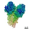 11835MC M: map data used to model this data C: citing same article ( |
|---|---|
| Similar structure data |


-Links
 PDBj
PDBj


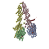
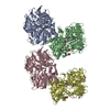
- Assembly
| Deposited unit | 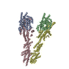
|
|---|---|
| 1 |
|
-Components
| #1: Protein | Mass: 56532.543 Da / Num. of mol.: 2 Source method: isolated from a genetically manipulated source Source: (gene. exp.) Candida albicans (yeast) / Gene: TUB4 / Production host:   Spodoptera frugiperda (fall armyworm) / References: UniProt: O93807 Spodoptera frugiperda (fall armyworm) / References: UniProt: O93807#2: Protein | | Mass: 101660.453 Da / Num. of mol.: 1 Source method: isolated from a genetically manipulated source Source: (gene. exp.) Candida albicans (yeast) / Gene: CAALFM_CR06740WA, orf19.708 / Production host: Spodoptera frugiperda (fall armyworm) / References: UniProt: Q59PZ2#3: Protein | | Mass: 92296.180 Da / Num. of mol.: 1 Source method: isolated from a genetically manipulated source Source: (gene. exp.) Candida albicans (yeast) / Gene: SPC98, CAALFM_CR01990CA, orf19.2600 / Production host: Spodoptera frugiperda (fall armyworm) / References: UniProt: A0A1D8PS42 |
|---|
-Experimental details
-Experiment
| Experiment | Method: ELECTRON MICROSCOPY |
|---|---|
| EM experiment | Aggregation state: PARTICLE / 3D reconstruction method: single particle reconstruction |
- Sample preparation
| Component | Name: gamma-Tubulin Small Complex / Type: COMPLEX Details: Candida albicans gamma-Tubulin Small Complex expressed and purified from SF21 cells Entity ID: all / Source: RECOMBINANT | ||||||||||||||||||||||||
|---|---|---|---|---|---|---|---|---|---|---|---|---|---|---|---|---|---|---|---|---|---|---|---|---|---|
| Molecular weight | Value: 0.3067 MDa / Experimental value: NO | ||||||||||||||||||||||||
| Source (natural) | Organism: Candida albicans (yeast) | ||||||||||||||||||||||||
| Source (recombinant) | Organism: Spodoptera frugiperda (fall armyworm) | ||||||||||||||||||||||||
| Buffer solution | pH: 7.4 | ||||||||||||||||||||||||
| Buffer component |
| ||||||||||||||||||||||||
| Specimen | Embedding applied: NO / Shadowing applied: NO / Staining applied: NO / Vitrification applied: YES | ||||||||||||||||||||||||
| Specimen support | Details: Gatan Solarus 950 plasma cleaner / Grid material: COPPER / Grid mesh size: 300 divisions/in. / Grid type: Quantifoil R2/1 | ||||||||||||||||||||||||
| Vitrification | Instrument: FEI VITROBOT MARK IV / Cryogen name: ETHANE / Humidity: 75 % / Chamber temperature: 298 K |
- Electron microscopy imaging
| Experimental equipment |  Model: Titan Krios / Image courtesy: FEI Company |
|---|---|
| Microscopy | Model: FEI TITAN KRIOS |
| Electron gun | Electron source:  FIELD EMISSION GUN / Accelerating voltage: 300 kV / Illumination mode: FLOOD BEAM FIELD EMISSION GUN / Accelerating voltage: 300 kV / Illumination mode: FLOOD BEAM |
| Electron lens | Mode: BRIGHT FIELD / Nominal defocus max: 3500 nm / Nominal defocus min: 2000 nm / Cs: 2.7 mm / Alignment procedure: COMA FREE |
| Specimen holder | Cryogen: NITROGEN / Specimen holder model: FEI TITAN KRIOS AUTOGRID HOLDER |
| Image recording | Electron dose: 2.1 e/Å2 / Detector mode: COUNTING / Film or detector model: GATAN K2 SUMMIT (4k x 4k) / Num. of grids imaged: 2 / Num. of real images: 1399 |
| EM imaging optics | Energyfilter slit width: 20 eV |
| Image scans | Width: 3710 / Height: 3838 / Movie frames/image: 20 |
- Processing
| Software | Name: PHENIX / Version: dev_3699: / Classification: refinement | |||||||||||||||||||||||||||||||||||||||||||||||||||||||
|---|---|---|---|---|---|---|---|---|---|---|---|---|---|---|---|---|---|---|---|---|---|---|---|---|---|---|---|---|---|---|---|---|---|---|---|---|---|---|---|---|---|---|---|---|---|---|---|---|---|---|---|---|---|---|---|---|
| EM software |
| |||||||||||||||||||||||||||||||||||||||||||||||||||||||
| CTF correction | Type: PHASE FLIPPING AND AMPLITUDE CORRECTION | |||||||||||||||||||||||||||||||||||||||||||||||||||||||
| Particle selection | Num. of particles selected: 1860000 | |||||||||||||||||||||||||||||||||||||||||||||||||||||||
| Symmetry | Point symmetry: C1 (asymmetric) | |||||||||||||||||||||||||||||||||||||||||||||||||||||||
| 3D reconstruction | Resolution: 3.6 Å / Resolution method: FSC 0.143 CUT-OFF / Num. of particles: 274326 / Symmetry type: POINT | |||||||||||||||||||||||||||||||||||||||||||||||||||||||
| Atomic model building |
| |||||||||||||||||||||||||||||||||||||||||||||||||||||||
| Refine LS restraints |
|