 Movie
Movie Controller

[English] 日本語
 Yorodumi
- PDB-7anv: Mutational and structural analysis of an ancestral D-type dye dec... -
+ Open data

- Basic information
| Entry | Database: PDB / ID: 7anv | ||||||
|---|---|---|---|---|---|---|---|
| Title | Mutational and structural analysis of an ancestral D-type dye decolorizing peroxidase | ||||||
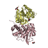
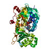
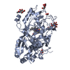
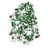
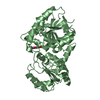
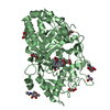
 Components Components | ancestral D-type dye decolorizing peroxidase | ||||||
 Keywords Keywords | OXIDOREDUCTASE / Dye decolorizing peroxidase / D-type DyP | ||||||
| Function / homology | PROTOPORPHYRIN IX CONTAINING FE / OXYGEN MOLECULE Function and homology information Function and homology information | ||||||
| Biological species |  Geotrichum candidum (yeast) Geotrichum candidum (yeast) | ||||||
| Method |  X-RAY DIFFRACTION / SYNCHROTRON / MOLECULAR REPLACEMENT / Resolution: 1.65 Å X-RAY DIFFRACTION / SYNCHROTRON / MOLECULAR REPLACEMENT / Resolution: 1.65 Å | ||||||
 Authors Authors | Rozeboom, H.J. / Fraaije, M.W. | ||||||
 Citation Citation | Journal: Febs J. / Year: 2021 Title: Mutational and structural analysis of an ancestral fungal dye-decolorizing peroxidase. Authors: Zitare, U.A. / Habib, M.H. / Rozeboom, H. / Mascotti, M.L. / Todorovic, S. / Fraaije, M.W. | ||||||
| History |
|
- Structure visualization
| Structure viewer | Molecule: MolmilJmol/JSmol |
|---|
- Downloads & links
-Download
| PDBx/mmCIF format | 7anv.cif.gz | 203.9 KB | Display | PDBx/mmCIF format |
|---|---|---|---|---|
| PDB format | pdb7anv.ent.gz | 159.9 KB | Display | PDB format |
| PDBx/mmJSON format | 7anv.json.gz | Tree view | PDBx/mmJSON format | |
| Others |  Other downloads Other downloads |
-Validation report
| Arichive directory | https://data.pdbj.org/pub/pdb/validation_reports/an/7anvftp://data.pdbj.org/pub/pdb/validation_reports/an/7anv | HTTPS FTP |
|---|
-Related structure data
| Related structure data |  6fskS S: Starting model for refinement |
|---|---|
| Similar structure data |


-Links
 PDBj
PDBj


- Assembly
| Deposited unit | 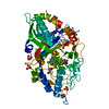
| ||||||||
|---|---|---|---|---|---|---|---|---|---|
| 1 |
| ||||||||
| Unit cell |
|
-Components
-Protein , 1 types, 1 molecules A
| #1: Protein | Mass: 53323.473 Da / Num. of mol.: 1 / Source method: obtained synthetically / Source: (synth.) Geotrichum candidum (yeast) |
|---|
-Non-polymers , 5 types, 408 molecules 


| #2: Chemical | ChemComp-HEM /  Mass: 616.487 Da / Num. of mol.: 1 / Source method: obtained synthetically / Formula: C34H32FeN4O4 Mass: 616.487 Da / Num. of mol.: 1 / Source method: obtained synthetically / Formula: C34H32FeN4O4 | ||||
|---|---|---|---|---|---|
| #3: Chemical | ChemComp-GOL /  Mass: 92.094 Da / Num. of mol.: 1 / Source method: obtained synthetically / Formula: C3H8O3 Mass: 92.094 Da / Num. of mol.: 1 / Source method: obtained synthetically / Formula: C3H8O3 | ||||
| #4: Chemical | ChemComp-SO4 /  Mass: 96.063 Da / Num. of mol.: 7 / Source method: obtained synthetically / Formula: SO4 Mass: 96.063 Da / Num. of mol.: 7 / Source method: obtained synthetically / Formula: SO4#5: Chemical | ChemComp-OXY / |  Mass: 31.999 Da / Num. of mol.: 1 / Source method: obtained synthetically / Formula: O2 Mass: 31.999 Da / Num. of mol.: 1 / Source method: obtained synthetically / Formula: O2#6: Water | ChemComp-HOH / | Mass: 18.015 Da / Num. of mol.: 398 / Source method: isolated from a natural source / Formula: H2O |
-Details
| Has ligand of interest | N |
|---|
-Experimental details
-Experiment
| Experiment | Method: X-RAY DIFFRACTION / Number of used crystals: 1 |
|---|
- Sample preparation
| Crystal | Density Matthews: 2.1 Å3/Da / Density % sol: 42 % |
|---|---|
| Crystal grow | Temperature: 294 K / Method: vapor diffusion, sitting drop / pH: 6.5 / Details: 2 M Ammonium sulfate, 0.1 M MES |
-Data collection
| Diffraction | Mean temperature: 100 K / Serial crystal experiment: N |
|---|---|
| Diffraction source | Source: SYNCHROTRON / Site: Diamond  / Beamline: I03 / Wavelength: 0.97625 Å / Beamline: I03 / Wavelength: 0.97625 Å |
| Detector | Type: DECTRIS EIGER2 XE 16M / Detector: PIXEL / Date: Jul 14, 2020 |
| Radiation | Protocol: SINGLE WAVELENGTH / Monochromatic (M) / Laue (L): M / Scattering type: x-ray |
| Radiation wavelength | Wavelength: 0.97625 Å / Relative weight: 1 |
| Reflection | Resolution: 1.65→59.15 Å / Num. obs: 55396 / % possible obs: 97 % / Redundancy: 10.3 % / CC1/2: 0.999 / Rmerge(I) obs: 0.065 / Rpim(I) all: 0.02 / Rrim(I) all: 0.068 / Net I/σ(I): 18.2 / Num. measured all: 570286 / Scaling rejects: 1012 |
| Reflection shell | Resolution: 1.65→1.68 Å / Redundancy: 3.9 % / Rmerge(I) obs: 0.759 / Num. unique obs: 2057 / CC1/2: 0.682 / Rpim(I) all: 0.422 / Rrim(I) all: 0.874 / % possible all: 75.2 |
- Processing
| Software |
| ||||||||||||||||||||||||||||||||||||||||||||||||||||||||||||||||||||||||||||||||||||||||||||||||||||||||||||||||||||||||||||||||||||||||||||||||||||||
|---|---|---|---|---|---|---|---|---|---|---|---|---|---|---|---|---|---|---|---|---|---|---|---|---|---|---|---|---|---|---|---|---|---|---|---|---|---|---|---|---|---|---|---|---|---|---|---|---|---|---|---|---|---|---|---|---|---|---|---|---|---|---|---|---|---|---|---|---|---|---|---|---|---|---|---|---|---|---|---|---|---|---|---|---|---|---|---|---|---|---|---|---|---|---|---|---|---|---|---|---|---|---|---|---|---|---|---|---|---|---|---|---|---|---|---|---|---|---|---|---|---|---|---|---|---|---|---|---|---|---|---|---|---|---|---|---|---|---|---|---|---|---|---|---|---|---|---|---|---|---|---|
| Refinement | Method to determine structure: MOLECULAR REPLACEMENT Starting model: 6FSK Resolution: 1.65→59.15 Å / Cor.coef. Fo:Fc: 0.971 / Cor.coef. Fo:Fc free: 0.958 / SU B: 3.669 / SU ML: 0.065 / Cross valid method: THROUGHOUT / σ(F): 0 / ESU R: 0.092 / ESU R Free: 0.093 / Stereochemistry target values: MAXIMUM LIKELIHOOD Details: U VALUES : WITH TLS ADDED HYDROGENS HAVE BEEN ADDED IN THE RIDING POSITIONS
| ||||||||||||||||||||||||||||||||||||||||||||||||||||||||||||||||||||||||||||||||||||||||||||||||||||||||||||||||||||||||||||||||||||||||||||||||||||||
| Solvent computation | Ion probe radii: 0.8 Å / Shrinkage radii: 0.8 Å / VDW probe radii: 1.2 Å / Solvent model: MASK | ||||||||||||||||||||||||||||||||||||||||||||||||||||||||||||||||||||||||||||||||||||||||||||||||||||||||||||||||||||||||||||||||||||||||||||||||||||||
| Displacement parameters | Biso max: 96.62 Å2 / Biso mean: 27.675 Å2 / Biso min: 14.43 Å2
| ||||||||||||||||||||||||||||||||||||||||||||||||||||||||||||||||||||||||||||||||||||||||||||||||||||||||||||||||||||||||||||||||||||||||||||||||||||||
| Refinement step | Cycle: final / Resolution: 1.65→59.15 Å
| ||||||||||||||||||||||||||||||||||||||||||||||||||||||||||||||||||||||||||||||||||||||||||||||||||||||||||||||||||||||||||||||||||||||||||||||||||||||
| Refine LS restraints |
| ||||||||||||||||||||||||||||||||||||||||||||||||||||||||||||||||||||||||||||||||||||||||||||||||||||||||||||||||||||||||||||||||||||||||||||||||||||||
| LS refinement shell | Resolution: 1.65→1.693 Å / Rfactor Rfree error: 0 / Total num. of bins used: 20
| ||||||||||||||||||||||||||||||||||||||||||||||||||||||||||||||||||||||||||||||||||||||||||||||||||||||||||||||||||||||||||||||||||||||||||||||||||||||
| Refinement TLS params. | Method: refined / Refine-ID: X-RAY DIFFRACTION
| ||||||||||||||||||||||||||||||||||||||||||||||||||||||||||||||||||||||||||||||||||||||||||||||||||||||||||||||||||||||||||||||||||||||||||||||||||||||
| Refinement TLS group |
|
