 Movie
Movie Controller


+ Open data

- Basic information
| Entry | Database: PDB / ID: 7anh | ||||||
|---|---|---|---|---|---|---|---|
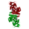
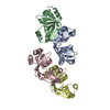
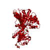
| Title | DdhaC | ||||||
 Components Components | GDP-L-fucose synthase | ||||||
 Keywords Keywords | SUGAR BINDING PROTEIN / reducatase / sugar nucleotide / sdr fold / enzyme | ||||||
| Function / homology |  Function and homology information Function and homology informationGDP-L-fucose synthase / GDP-L-fucose synthase activity / 'de novo' GDP-L-fucose biosynthetic process / NADP+ binding / isomerase activity Similarity search - Function | ||||||
| Biological species |   Campylobacter jejuni (Campylobacter) Campylobacter jejuni (Campylobacter) | ||||||
| Method |  X-RAY DIFFRACTION / MOLECULAR REPLACEMENT / Resolution: 2.08 Å X-RAY DIFFRACTION / MOLECULAR REPLACEMENT / Resolution: 2.08 Å | ||||||
 Authors Authors | Naismith, J.H. / Woodward, L. | ||||||
| Funding support |  United Kingdom, 1items United Kingdom, 1items
| ||||||
 Citation Citation | Journal: To Be Published Title: DdhaC Authors: Naismith, J.H. / Woodward, L. | ||||||
| History |
|
- Structure visualization
| Structure viewer | Molecule: MolmilJmol/JSmol |
|---|
- Downloads & links
-Download
| PDBx/mmCIF format | 7anh.cif.gz | 83.8 KB | Display | PDBx/mmCIF format |
|---|---|---|---|---|
| PDB format | pdb7anh.ent.gz | 63 KB | Display | PDB format |
| PDBx/mmJSON format | 7anh.json.gz | Tree view | PDBx/mmJSON format | |
| Others |  Other downloads Other downloads |
-Validation report
| Summary document | 7anh_validation.pdf.gz | 421.4 KB | Display | wwPDB validaton report |
|---|---|---|---|---|
| Full document | 7anh_full_validation.pdf.gz | 423.6 KB | Display | |
| Data in XML | 7anh_validation.xml.gz | 14.4 KB | Display | |
| Data in CIF | 7anh_validation.cif.gz | 20 KB | Display | |
| Arichive directory | https://data.pdbj.org/pub/pdb/validation_reports/an/7anhftp://data.pdbj.org/pub/pdb/validation_reports/an/7anh | HTTPS FTP |
-Related structure data
| Related structure data |  1bsvS S: Starting model for refinement |
|---|---|
| Similar structure data |


-Links
 PDBj
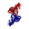
PDBj- Assembly
| Deposited unit | 
| ||||||||
|---|---|---|---|---|---|---|---|---|---|
| 1 | 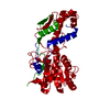
| ||||||||
| Unit cell |
| ||||||||
| Components on special symmetry positions |
|
-Components
| #1: Protein | Mass: 40521.664 Da / Num. of mol.: 1 Source method: isolated from a genetically manipulated source Source: (gene. exp.) Campylobacter jejuni (Campylobacter)Gene: AY595_07840, B9Q63_05455, BB943_08725, D0W34_08630, D5I02_08530, DK813_08295, DQX79_06120, DW530_02880, F1P94_08750, FQZ47_07910, FRQ83_08390, FW031_08555, GAX04_08405, GRS20_08805, GSH24_ ...Gene: AY595_07840, B9Q63_05455, BB943_08725, D0W34_08630, D5I02_08530, DK813_08295, DQX79_06120, DW530_02880, F1P94_08750, FQZ47_07910, FRQ83_08390, FW031_08555, GAX04_08405, GRS20_08805, GSH24_04185, GY415_001379, GZD82_001462 Production host: |
|---|---|
| #2: Water | ChemComp-HOH /  Mass: 18.015 Da / Num. of mol.: 87 / Source method: isolated from a natural source / Formula: H2O Mass: 18.015 Da / Num. of mol.: 87 / Source method: isolated from a natural source / Formula: H2O |
-Experimental details
-Experiment
| Experiment | Method: X-RAY DIFFRACTION / Number of used crystals: 1 |
|---|
- Sample preparation
| Crystal | Density Matthews: 2.69 Å3/Da / Density % sol: 54.25 % |
|---|---|
| Crystal grow | Temperature: 298 K / Method: vapor diffusion, sitting drop Details: 54 % (w/v) PEG 400, 0.1 M HEPES pH 7.5, 0.08 M ammonium citrate |
-Data collection
| Diffraction | Mean temperature: 120 K / Serial crystal experiment: N |
|---|---|
| Diffraction source | Source: ROTATING ANODE / Type: RIGAKU MICROMAX-007 HF / Wavelength: 1.5418 Å |
| Detector | Type: RIGAKU SATURN 944 / Detector: CCD / Date: Oct 4, 2016 |
| Radiation | Protocol: SINGLE WAVELENGTH / Monochromatic (M) / Laue (L): M / Scattering type: x-ray |
| Radiation wavelength | Wavelength: 1.5418 Å / Relative weight: 1 |
| Reflection | Resolution: 2.08→99 Å / Num. obs: 25782 / % possible obs: 100 % / Redundancy: 12.5 % / CC1/2: 0.9 / Rmerge(I) obs: 0.163 / Net I/σ(I): 11.5 |
| Reflection shell | Resolution: 2.08→2.14 Å / Rmerge(I) obs: 0.61 / Mean I/σ(I) obs: 3.3 / Num. unique obs: 1861 / CC1/2: 0.8 |
- Processing
| Software |
| ||||||||||||||||||||||||||||||||||||||||||||||||||||||||||||
|---|---|---|---|---|---|---|---|---|---|---|---|---|---|---|---|---|---|---|---|---|---|---|---|---|---|---|---|---|---|---|---|---|---|---|---|---|---|---|---|---|---|---|---|---|---|---|---|---|---|---|---|---|---|---|---|---|---|---|---|---|---|
| Refinement | Method to determine structure: MOLECULAR REPLACEMENT Starting model: 1BSV Resolution: 2.08→93.2 Å / Cor.coef. Fo:Fc: 0.94 / Cor.coef. Fo:Fc free: 0.915 / SU B: 4.546 / SU ML: 0.12 / Cross valid method: THROUGHOUT / σ(F): 0 / ESU R: 0.182 / ESU R Free: 0.165 / Stereochemistry target values: MAXIMUM LIKELIHOOD Details: HYDROGENS HAVE BEEN ADDED IN THE RIDING POSITIONS U VALUES : REFINED INDIVIDUALLY
| ||||||||||||||||||||||||||||||||||||||||||||||||||||||||||||
| Solvent computation | Ion probe radii: 0.8 Å / Shrinkage radii: 0.8 Å / VDW probe radii: 1.2 Å / Solvent model: MASK | ||||||||||||||||||||||||||||||||||||||||||||||||||||||||||||
| Displacement parameters | Biso max: 91.53 Å2 / Biso mean: 26.362 Å2 / Biso min: 8.2 Å2
| ||||||||||||||||||||||||||||||||||||||||||||||||||||||||||||
| Refinement step | Cycle: final / Resolution: 2.08→93.2 Å
| ||||||||||||||||||||||||||||||||||||||||||||||||||||||||||||
| Refine LS restraints |
| ||||||||||||||||||||||||||||||||||||||||||||||||||||||||||||
| LS refinement shell | Resolution: 2.08→2.133 Å / Rfactor Rfree error: 0
|
