 Movie
Movie Controller


+ Open data

- Basic information
| Entry | Database: PDB / ID: 7amk | ||||||
|---|---|---|---|---|---|---|---|
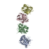
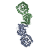
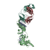
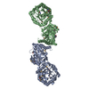
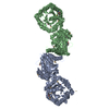
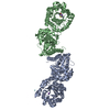
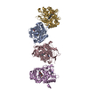
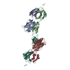
| Title | Zebrafish RET Cadherin Like Domains 1 to 4. | ||||||
 Components Components | Proto-oncogene tyrosine-protein kinase receptor Ret | ||||||
 Keywords Keywords | SIGNALING PROTEIN / Ligand recognition / receptor tyrosine kinase / glycosylation | ||||||
| Function / homology |  Function and homology information Function and homology informationbranchiomeric skeletal muscle development / pronephros morphogenesis / RAF/MAP kinase cascade / RET signaling / neural crest cell migration involved in autonomic nervous system development / enteric nervous system development / homophilic cell-cell adhesion / transmembrane receptor protein tyrosine kinase activity / endomembrane system / cell surface receptor protein tyrosine kinase signaling pathway ...branchiomeric skeletal muscle development / pronephros morphogenesis / RAF/MAP kinase cascade / RET signaling / neural crest cell migration involved in autonomic nervous system development / enteric nervous system development / homophilic cell-cell adhesion / transmembrane receptor protein tyrosine kinase activity / endomembrane system / cell surface receptor protein tyrosine kinase signaling pathway / receptor protein-tyrosine kinase / neuron projection development / protein-containing complex assembly / signaling receptor complex / axon / calcium ion binding / protein homodimerization activity / ATP binding / plasma membrane Similarity search - Function | ||||||
| Biological species |  | ||||||
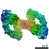
| Method |  X-RAY DIFFRACTION / SYNCHROTRON / MOLECULAR REPLACEMENT / Resolution: 2.2 Å X-RAY DIFFRACTION / SYNCHROTRON / MOLECULAR REPLACEMENT / Resolution: 2.2 Å | ||||||
 Authors Authors | Purkiss, A.G. / McDonald, N.Q. / Goodman, K.M. / Narowtek, A. / Knowles, P.P. | ||||||
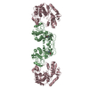
 Citation Citation | Journal: Structure / Year: 2021 Title: A two-site flexible clamp mechanism for RET-GDNF-GFRα1 assembly reveals both conformational adaptation and strict geometric spacing. Authors: Sarah E Adams / Andrew G Purkiss / Phillip P Knowles / Andrea Nans / David C Briggs / Annabel Borg / Christopher P Earl / Kerry M Goodman / Agata Nawrotek / Aaron J Borg / Pauline B McIntosh ...Authors: Sarah E Adams / Andrew G Purkiss / Phillip P Knowles / Andrea Nans / David C Briggs / Annabel Borg / Christopher P Earl / Kerry M Goodman / Agata Nawrotek / Aaron J Borg / Pauline B McIntosh / Francesca M Houghton / Svend Kjær / Neil Q McDonald /  Abstract: RET receptor tyrosine kinase plays vital developmental and neuroprotective roles in metazoans. GDNF family ligands (GFLs) when bound to cognate GFRα co-receptors recognize and activate RET ...RET receptor tyrosine kinase plays vital developmental and neuroprotective roles in metazoans. GDNF family ligands (GFLs) when bound to cognate GFRα co-receptors recognize and activate RET stimulating its cytoplasmic kinase function. The principles for RET ligand-co-receptor recognition are incompletely understood. Here, we report a crystal structure of the cadherin-like module (CLD1-4) from zebrafish RET revealing interdomain flexibility between CLD2 and CLD3. Comparison with a cryo-electron microscopy structure of a ligand-engaged zebrafish RET-GDNF-GFRα1a complex indicates conformational changes within a clade-specific CLD3 loop adjacent to the co-receptor. Our observations indicate that RET is a molecular clamp with a flexible calcium-dependent arm that adapts to different GFRα co-receptors, while its rigid arm recognizes a GFL dimer to align both membrane-proximal cysteine-rich domains. We also visualize linear arrays of RET-GDNF-GFRα1a suggesting that a conserved contact stabilizes higher-order species. Our study reveals that ligand-co-receptor recognition by RET involves both receptor plasticity and strict spacing of receptor dimers by GFL ligands. | ||||||
| History |
|
- Structure visualization
| Structure viewer | Molecule: MolmilJmol/JSmol |
|---|
- Downloads & links
-Download
| PDBx/mmCIF format | 7amk.cif.gz | 496.3 KB | Display | PDBx/mmCIF format |
|---|---|---|---|---|
| PDB format | pdb7amk.ent.gz | 339.9 KB | Display | PDB format |
| PDBx/mmJSON format | 7amk.json.gz | Tree view | PDBx/mmJSON format | |
| Others |  Other downloads Other downloads |
-Validation report
| Arichive directory | https://data.pdbj.org/pub/pdb/validation_reports/am/7amkftp://data.pdbj.org/pub/pdb/validation_reports/am/7amk | HTTPS FTP |
|---|
-Related structure data
| Related structure data |  7ab8C 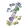 7amlC  2x2uS C: citing same article ( S: Starting model for refinement |
|---|---|
| Similar structure data |


-Links
 PDBj
PDBj
- Assembly
| Deposited unit | 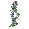
| ||||||||||||
|---|---|---|---|---|---|---|---|---|---|---|---|---|---|
| 1 |
| ||||||||||||
| Unit cell |
| ||||||||||||
| Noncrystallographic symmetry (NCS) | NCS oper: (Code: givenMatrix: (0.430793954191, -0.900333557273, -0.0617742234261), (-0.902265964183, -0.431079226797, -0.00931826708919), (-0.0182400359145, 0.0597510323868, -0.998046649821)Vector: ...NCS oper: (Code: given Matrix: (0.430793954191, -0.900333557273, -0.0617742234261), Vector: |
-Components
-Protein , 1 types, 2 molecules AB
| #1: Protein | Mass: 54610.270 Da / Num. of mol.: 2 Source method: isolated from a genetically manipulated source Source: (gene. exp.)  unidentified baculovirus unidentified baculovirusReferences: UniProt: A8E7C6, receptor protein-tyrosine kinase |
|---|
-Sugars , 7 types, 12 molecules 
| #2: Polysaccharide | alpha-D-mannopyranose-(1-6)-alpha-D-mannopyranose-(1-3)-[alpha-D-mannopyranose-(1-6)]beta-D- ...alpha-D-mannopyranose-(1-6)-alpha-D-mannopyranose-(1-3)-[alpha-D-mannopyranose-(1-6)]beta-D-mannopyranose-(1-4)-2-acetamido-2-deoxy-beta-D-glucopyranose-(1-4)-[alpha-L-fucopyranose-(1-6)]2-acetamido-2-deoxy-beta-D-glucopyranose Source method: isolated from a genetically manipulated source | ||||||||||
|---|---|---|---|---|---|---|---|---|---|---|---|
| #3: Polysaccharide | Source method: isolated from a genetically manipulated source #4: Polysaccharide | Source method: isolated from a genetically manipulated source #5: Polysaccharide | alpha-D-mannopyranose-(1-6)-beta-D-mannopyranose-(1-4)-2-acetamido-2-deoxy-beta-D-glucopyranose-(1- ...alpha-D-mannopyranose-(1-6)-beta-D-mannopyranose-(1-4)-2-acetamido-2-deoxy-beta-D-glucopyranose-(1-4)-[alpha-L-fucopyranose-(1-6)]2-acetamido-2-deoxy-beta-D-glucopyranose | Source method: isolated from a genetically manipulated source #6: Polysaccharide | beta-D-mannopyranose-(1-4)-2-acetamido-2-deoxy-beta-D-glucopyranose-(1-4)-[alpha-L-fucopyranose-(1- ...beta-D-mannopyranose-(1-4)-2-acetamido-2-deoxy-beta-D-glucopyranose-(1-4)-[alpha-L-fucopyranose-(1-6)]2-acetamido-2-deoxy-beta-D-glucopyranose | Source method: isolated from a genetically manipulated source #7: Polysaccharide | alpha-D-mannopyranose-(1-3)-beta-D-mannopyranose-(1-4)-2-acetamido-2-deoxy-beta-D-glucopyranose-(1- ...alpha-D-mannopyranose-(1-3)-beta-D-mannopyranose-(1-4)-2-acetamido-2-deoxy-beta-D-glucopyranose-(1-4)-[alpha-L-fucopyranose-(1-6)]2-acetamido-2-deoxy-beta-D-glucopyranose | Source method: isolated from a genetically manipulated source #9: Sugar |  Type: D-saccharide, beta linking / Mass: 221.208 Da / Num. of mol.: 3 Type: D-saccharide, beta linking / Mass: 221.208 Da / Num. of mol.: 3Source method: isolated from a genetically manipulated source Formula: C8H15NO6 |
-Non-polymers , 6 types, 245 molecules 


| #8: Chemical | ChemComp-CA /  Mass: 40.078 Da / Num. of mol.: 6 Mass: 40.078 Da / Num. of mol.: 6Source method: isolated from a genetically manipulated source Formula: Ca #10: Chemical | ChemComp-GOL /  Mass: 92.094 Da / Num. of mol.: 4 Mass: 92.094 Da / Num. of mol.: 4Source method: isolated from a genetically manipulated source Formula: C3H8O3 #11: Chemical | ChemComp-PGE /  Mass: 150.173 Da / Num. of mol.: 5 Mass: 150.173 Da / Num. of mol.: 5Source method: isolated from a genetically manipulated source Formula: C6H14O4 #12: Chemical | ChemComp-PEG /  Mass: 106.120 Da / Num. of mol.: 10 Mass: 106.120 Da / Num. of mol.: 10Source method: isolated from a genetically manipulated source Formula: C4H10O3 #13: Chemical | ChemComp-MES / |  Mass: 195.237 Da / Num. of mol.: 1 Mass: 195.237 Da / Num. of mol.: 1Source method: isolated from a genetically manipulated source Formula: C6H13NO4S / Comment: pH buffer*YM #14: Water | ChemComp-HOH / | Mass: 18.015 Da / Num. of mol.: 219 / Source method: isolated from a natural source / Formula: H2O |
|---|
-Details
| Has ligand of interest | N |
|---|---|
| Has protein modification | Y |
-Experimental details
-Experiment
| Experiment | Method: X-RAY DIFFRACTION / Number of used crystals: 1 |
|---|
- Sample preparation
| Crystal | Density Matthews: 3.28 Å3/Da / Density % sol: 62.53 % |
|---|---|
| Crystal grow | Temperature: 293 K / Method: vapor diffusion, sitting drop / pH: 6.2 / Details: 50 mM MES (pH 6.2), 31.5 % PEG MME 350 (v/v) |
-Data collection
| Diffraction | Mean temperature: 100 K / Serial crystal experiment: N |
|---|---|
| Diffraction source | Source: SYNCHROTRON / Site: Diamond / Beamline: I03 / Wavelength: 0.9787 Å |
| Detector | Type: DECTRIS PILATUS 6M / Detector: PIXEL / Date: May 24, 2015 / Details: Mirrors |
| Radiation | Protocol: SINGLE WAVELENGTH / Monochromatic (M) / Laue (L): M / Scattering type: x-ray |
| Radiation wavelength | Wavelength: 0.9787 Å / Relative weight: 1 |
| Reflection | Resolution: 2.08→65.96 Å / Num. obs: 79868 / % possible obs: 98.2 % / Redundancy: 3.4 % / Biso Wilson estimate: 28.87 Å2 / CC1/2: 0.996 / Rmerge(I) obs: 0.08 / Rpim(I) all: 0.051 / Rrim(I) all: 0.095 / Net I/σ(I): 6.2 |
| Reflection shell | Resolution: 2.08→2.19 Å / Redundancy: 3.3 % / Rmerge(I) obs: 1.186 / Mean I/σ(I) obs: 1.4 / Num. unique obs: 11549 / CC1/2: 0.58 / Rpim(I) all: 0.765 / Rrim(I) all: 1.416 / % possible all: 97.2 |
- Processing
| Software |
| |||||||||||||||||||||||||||||||||||||||||||||||||||||||||||||||||||||||||||||||||||||||||||||||||||||||||||||||||||||||||||||||||||||||||||||||||||||||||||||||||||||||||||||||
|---|---|---|---|---|---|---|---|---|---|---|---|---|---|---|---|---|---|---|---|---|---|---|---|---|---|---|---|---|---|---|---|---|---|---|---|---|---|---|---|---|---|---|---|---|---|---|---|---|---|---|---|---|---|---|---|---|---|---|---|---|---|---|---|---|---|---|---|---|---|---|---|---|---|---|---|---|---|---|---|---|---|---|---|---|---|---|---|---|---|---|---|---|---|---|---|---|---|---|---|---|---|---|---|---|---|---|---|---|---|---|---|---|---|---|---|---|---|---|---|---|---|---|---|---|---|---|---|---|---|---|---|---|---|---|---|---|---|---|---|---|---|---|---|---|---|---|---|---|---|---|---|---|---|---|---|---|---|---|---|---|---|---|---|---|---|---|---|---|---|---|---|---|---|---|---|---|
| Refinement | Method to determine structure: MOLECULAR REPLACEMENT Starting model: 2x2u Resolution: 2.2→65.96 Å / SU ML: 0.3521 / Cross valid method: FREE R-VALUE / σ(F): 1.96 / Phase error: 29.5782 Stereochemistry target values: GeoStd + Monomer Library + CDL v1.2
| |||||||||||||||||||||||||||||||||||||||||||||||||||||||||||||||||||||||||||||||||||||||||||||||||||||||||||||||||||||||||||||||||||||||||||||||||||||||||||||||||||||||||||||||
| Solvent computation | Shrinkage radii: 0.9 Å / VDW probe radii: 1.11 Å / Solvent model: FLAT BULK SOLVENT MODEL | |||||||||||||||||||||||||||||||||||||||||||||||||||||||||||||||||||||||||||||||||||||||||||||||||||||||||||||||||||||||||||||||||||||||||||||||||||||||||||||||||||||||||||||||
| Displacement parameters | Biso mean: 39.39 Å2 | |||||||||||||||||||||||||||||||||||||||||||||||||||||||||||||||||||||||||||||||||||||||||||||||||||||||||||||||||||||||||||||||||||||||||||||||||||||||||||||||||||||||||||||||
| Refinement step | Cycle: LAST / Resolution: 2.2→65.96 Å
| |||||||||||||||||||||||||||||||||||||||||||||||||||||||||||||||||||||||||||||||||||||||||||||||||||||||||||||||||||||||||||||||||||||||||||||||||||||||||||||||||||||||||||||||
| Refine LS restraints |
| |||||||||||||||||||||||||||||||||||||||||||||||||||||||||||||||||||||||||||||||||||||||||||||||||||||||||||||||||||||||||||||||||||||||||||||||||||||||||||||||||||||||||||||||
| LS refinement shell |
| |||||||||||||||||||||||||||||||||||||||||||||||||||||||||||||||||||||||||||||||||||||||||||||||||||||||||||||||||||||||||||||||||||||||||||||||||||||||||||||||||||||||||||||||
| Refinement TLS params. | Method: refined / Refine-ID: X-RAY DIFFRACTION
| |||||||||||||||||||||||||||||||||||||||||||||||||||||||||||||||||||||||||||||||||||||||||||||||||||||||||||||||||||||||||||||||||||||||||||||||||||||||||||||||||||||||||||||||
| Refinement TLS group | Refine-ID: X-RAY DIFFRACTION
|