 Movie
Movie Controller


+ Open data

- Basic information
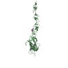
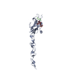
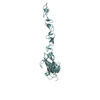
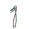
| Entry | Database: PDB / ID: 7alt | ||||||||||||
|---|---|---|---|---|---|---|---|---|---|---|---|---|---|
| Title | Structure of Drosophila Serrate C2-DSL-EGF1-EGF2 | ||||||||||||
 Components Components | Protein serrate | ||||||||||||
 Keywords Keywords | SIGNALING PROTEIN / Serrate Notch Ligand C2 / DSL and EGF domains | ||||||||||||
| Function / homology |  Function and homology information Function and homology informationPre-NOTCH Processing in Golgi / NOTCH4 Activation and Transmission of Signal to the Nucleus / Negative regulation of NOTCH4 signaling / eye-antennal disc development / crystal cell differentiation / negative regulation of lamellocyte differentiation / cuticle pattern formation / lateral inhibition / second mitotic wave involved in compound eye morphogenesis / imaginal disc-derived wing margin morphogenesis ...Pre-NOTCH Processing in Golgi / NOTCH4 Activation and Transmission of Signal to the Nucleus / Negative regulation of NOTCH4 signaling / eye-antennal disc development / crystal cell differentiation / negative regulation of lamellocyte differentiation / cuticle pattern formation / lateral inhibition / second mitotic wave involved in compound eye morphogenesis / imaginal disc-derived wing margin morphogenesis / imaginal disc-derived leg segmentation / imaginal disc-derived leg morphogenesis / wing disc anterior/posterior pattern formation / larval salivary gland morphogenesis / larval lymph gland hemopoiesis / compound eye development / imaginal disc-derived wing morphogenesis / glycosphingolipid binding / germ-line stem cell population maintenance / apical cortex / sensory organ development / Notch binding / axolemma / salivary gland morphogenesis / glial cell proliferation / Notch signaling pathway / apical plasma membrane / receptor ligand activity / calcium ion binding / cell surface / plasma membrane Similarity search - Function | ||||||||||||
| Biological species |  | ||||||||||||
| Method |  X-RAY DIFFRACTION / SYNCHROTRON / MOLECULAR REPLACEMENT / Resolution: 2.03 Å X-RAY DIFFRACTION / SYNCHROTRON / MOLECULAR REPLACEMENT / Resolution: 2.03 Å | ||||||||||||
 Authors Authors | Suckling, R. / Johnson, S. / Lea, S.M. | ||||||||||||
| Funding support |  United Kingdom, 3items United Kingdom, 3items
| ||||||||||||
 Citation Citation | Journal: Embo Rep. / Year: 2021 Title: The conserved C2 phospholipid-binding domain in Delta contributes to robust Notch signalling. Authors: Martins, T. / Meng, Y. / Korona, B. / Suckling, R. / Johnson, S. / Handford, P.A. / Lea, S.M. / Bray, S.J. | ||||||||||||
| History |
|
- Structure visualization
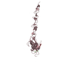
| Structure viewer | Molecule: MolmilJmol/JSmol |
|---|
- Downloads & links
-Download
| PDBx/mmCIF format | 7alt.cif.gz | 249.3 KB | Display | PDBx/mmCIF format |
|---|---|---|---|---|
| PDB format | pdb7alt.ent.gz | 163.6 KB | Display | PDB format |
| PDBx/mmJSON format | 7alt.json.gz | Tree view | PDBx/mmJSON format | |
| Others |  Other downloads Other downloads |
-Validation report
| Arichive directory | https://data.pdbj.org/pub/pdb/validation_reports/al/7altftp://data.pdbj.org/pub/pdb/validation_reports/al/7alt | HTTPS FTP |
|---|
-Related structure data
| Related structure data |  7aljC 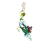 7alkC 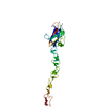 5mw7S S: Starting model for refinement C: citing same article ( |
|---|---|
| Similar structure data |


-Links
 PDBj
PDBj


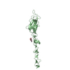
- Assembly
| Deposited unit | 
| |||||||||||||||||||||||||||||||||||||||||||||||||||||||||||||||||
|---|---|---|---|---|---|---|---|---|---|---|---|---|---|---|---|---|---|---|---|---|---|---|---|---|---|---|---|---|---|---|---|---|---|---|---|---|---|---|---|---|---|---|---|---|---|---|---|---|---|---|---|---|---|---|---|---|---|---|---|---|---|---|---|---|---|---|
| 1 | 
| |||||||||||||||||||||||||||||||||||||||||||||||||||||||||||||||||
| 2 | 
| |||||||||||||||||||||||||||||||||||||||||||||||||||||||||||||||||
| Unit cell |
| |||||||||||||||||||||||||||||||||||||||||||||||||||||||||||||||||
| Noncrystallographic symmetry (NCS) | NCS domain:
NCS domain segments:
NCS oper: (Code: givenMatrix: (-0.999906210829, -0.000599648350543, 0.0136824693301), (-0.000858325876441, 0.999820864457, -0.0189077304919), (-0.0136686803242, -0.0189177011691, -0.999727606781) ...NCS oper: (Code: given Matrix: (-0.999906210829, -0.000599648350543, 0.0136824693301), Vector: |
-Components
| #1: Protein | Mass: 29610.537 Da / Num. of mol.: 2 Source method: isolated from a genetically manipulated source Source: (gene. exp.) #2: Polysaccharide | 2-acetamido-2-deoxy-beta-D-glucopyranose-(1-4)-2-acetamido-2-deoxy-beta-D-glucopyranose | Source method: isolated from a genetically manipulated source #3: Chemical |   Mass: 40.078 Da / Num. of mol.: 2 / Source method: obtained synthetically / Formula: Ca Mass: 40.078 Da / Num. of mol.: 2 / Source method: obtained synthetically / Formula: Ca#4: Sugar |   Type: D-saccharide, beta linking / Mass: 221.208 Da / Num. of mol.: 2 / Source method: obtained synthetically / Formula: C8H15NO6 Type: D-saccharide, beta linking / Mass: 221.208 Da / Num. of mol.: 2 / Source method: obtained synthetically / Formula: C8H15NO6#5: Water | ChemComp-HOH / |  Mass: 18.015 Da / Num. of mol.: 118 / Source method: isolated from a natural source / Formula: H2O Mass: 18.015 Da / Num. of mol.: 118 / Source method: isolated from a natural source / Formula: H2OHas ligand of interest | N | Has protein modification | Y | |
|---|
-Experimental details
-Experiment
| Experiment | Method: X-RAY DIFFRACTION / Number of used crystals: 1 |
|---|
- Sample preparation
| Crystal | Density Matthews: 2.56 Å3/Da / Density % sol: 52.01 % |
|---|---|
| Crystal grow | Temperature: 290 K / Method: vapor diffusion, sitting drop / Details: 0.1M imidazole malate pH 7, 25% PEG4K |
-Data collection
| Diffraction | Mean temperature: 100 K / Serial crystal experiment: N |
|---|---|
| Diffraction source | Source: SYNCHROTRON / Site: Diamond / Beamline: I02 / Wavelength: 0.97949 Å |
| Detector | Type: DECTRIS EIGER X 16M / Detector: PIXEL / Date: May 4, 2015 |
| Radiation | Protocol: SINGLE WAVELENGTH / Monochromatic (M) / Laue (L): M / Scattering type: x-ray |
| Radiation wavelength | Wavelength: 0.97949 Å / Relative weight: 1 |
| Reflection | Resolution: 2.03→45.61 Å / Num. obs: 38729 / % possible obs: 98.89 % / Redundancy: 3.3 % / Biso Wilson estimate: 31.49 Å2 / CC1/2: 0.998 / Net I/σ(I): 11.1 |
| Reflection shell | Resolution: 2.03→2.103 Å / Num. unique obs: 3814 / CC1/2: 0.769 |
- Processing
| Software |
| |||||||||||||||||||||||||||||||||||||||||||||||||||||||||||||||||||||||||||||||||||||||||||||||||||||||||
|---|---|---|---|---|---|---|---|---|---|---|---|---|---|---|---|---|---|---|---|---|---|---|---|---|---|---|---|---|---|---|---|---|---|---|---|---|---|---|---|---|---|---|---|---|---|---|---|---|---|---|---|---|---|---|---|---|---|---|---|---|---|---|---|---|---|---|---|---|---|---|---|---|---|---|---|---|---|---|---|---|---|---|---|---|---|---|---|---|---|---|---|---|---|---|---|---|---|---|---|---|---|---|---|---|---|---|
| Refinement | Method to determine structure: MOLECULAR REPLACEMENT Starting model: 5MW7 Resolution: 2.03→45.61 Å / SU ML: 0.2152 / Cross valid method: FREE R-VALUE / σ(F): 1.34 / Phase error: 30.3516 Stereochemistry target values: GeoStd + Monomer Library + CDL v1.2
| |||||||||||||||||||||||||||||||||||||||||||||||||||||||||||||||||||||||||||||||||||||||||||||||||||||||||
| Solvent computation | Shrinkage radii: 0.9 Å / VDW probe radii: 1.11 Å / Solvent model: FLAT BULK SOLVENT MODEL | |||||||||||||||||||||||||||||||||||||||||||||||||||||||||||||||||||||||||||||||||||||||||||||||||||||||||
| Displacement parameters | Biso mean: 41.24 Å2 | |||||||||||||||||||||||||||||||||||||||||||||||||||||||||||||||||||||||||||||||||||||||||||||||||||||||||
| Refinement step | Cycle: LAST / Resolution: 2.03→45.61 Å
| |||||||||||||||||||||||||||||||||||||||||||||||||||||||||||||||||||||||||||||||||||||||||||||||||||||||||
| Refine LS restraints |
| |||||||||||||||||||||||||||||||||||||||||||||||||||||||||||||||||||||||||||||||||||||||||||||||||||||||||
| Refine LS restraints NCS | Type: Torsion NCS / Rms dev position: 2.92239358248 Å | |||||||||||||||||||||||||||||||||||||||||||||||||||||||||||||||||||||||||||||||||||||||||||||||||||||||||
| LS refinement shell |
|
