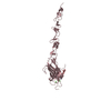
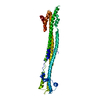
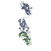
- PDB-1x79: Crystal structure of human GGA1 GAT domain complexed with the GAT... -
+
Open data
ID or keywords:
Loading...
-
Basic information
Entry
Database: PDB / ID: 1x79
Title
Crystal structure of human GGA1 GAT domain complexed with the GAT-binding domain of Rabaptin5
Components
ADP-ribosylation factor binding protein GGA1
Rab GTPase binding effector protein 1
Keywords
PROTEIN TRANSPORT / Rabaptin5 / GGA protein / GAT domain / intracellular trafficking
Function / homology
Function and homology information
protein localization to ciliary membrane / Golgi to plasma membrane transport / Golgi to plasma membrane protein transport / TBC/RABGAPs / retrograde transport, endosome to Golgi / protein localization to cell surface / regulation of postsynaptic neurotransmitter receptor internalization / endocytic vesicle / vesicle-mediated transport / phosphatidylinositol binding ...protein localization to ciliary membrane / Golgi to plasma membrane transport / Golgi to plasma membrane protein transport / TBC/RABGAPs / retrograde transport, endosome to Golgi / protein localization to cell surface / regulation of postsynaptic neurotransmitter receptor internalization / endocytic vesicle / vesicle-mediated transport / phosphatidylinositol binding / GTPase activator activity / growth factor activity / ubiquitin binding / trans-Golgi network / intracellular protein transport / recycling endosome / small GTPase binding / endocytosis / intracellular protein localization / protein transport / early endosome membrane / membrane fusion / early endosome / endosome / endosome membrane / Amyloid fiber formation / protein domain specific binding / apoptotic process / glutamatergic synapse / Golgi apparatus / protein homodimerization activity / protein-containing complex / membrane / cytosol Similarity search - Function
Rabaptin / Rabaptin, GTPase-Rab5 binding domain / Rabaptin coiled-coil domain / Rabaptin / Rabaptin-like protein / Methane Monooxygenase Hydroxylase; Chain G, domain 1 - #160 / ADP-ribosylation factor-binding protein GGA3 / N-terminal extension of GAT domain / N-terminal extension of GAT domain / GAT domain ...Rabaptin / Rabaptin, GTPase-Rab5 binding domain / Rabaptin coiled-coil domain / Rabaptin / Rabaptin-like protein / Methane Monooxygenase Hydroxylase; Chain G, domain 1 - #160 / ADP-ribosylation factor-binding protein GGA3 / N-terminal extension of GAT domain / N-terminal extension of GAT domain / GAT domain / GAT domain superfamily / GAT domain / GAT domain profile. / Single alpha-helices involved in coiled-coils or other helix-helix interfaces - #340 / VHS domain / VHS domain / VHS domain profile. / Domain present in VPS-27, Hrs and STAM / Gamma-adaptin ear (GAE) domain / Gamma-adaptin ear (GAE) domain profile. / Clathrin adaptor, alpha/beta/gamma-adaptin, appendage, Ig-like subdomain / Adaptin C-terminal domain / Adaptin C-terminal domain / Clathrin adaptor, appendage, Ig-like subdomain superfamily / ENTH/VHS / Single alpha-helices involved in coiled-coils or other helix-helix interfaces / Methane Monooxygenase Hydroxylase; Chain G, domain 1 / Up-down Bundle / Mainly Alpha Similarity search - Domain/homology
2,3-DIHYDROXY-1,4-DITHIOBUTANE / Rab GTPase-binding effector protein 1 / ADP-ribosylation factor-binding protein GGA1 Similarity search - Component
Mass: 18.015 Da / Num. of mol.: 117 / Source method: isolated from a natural source / Formula: H2O
-
Experimental details
-
Experiment
Experiment
Method: X-RAY DIFFRACTION / Number of used crystals: 1
-
Sample preparation
Crystal
Density Matthews: 5.4 Å3/Da / Density % sol: 77 % Description: The phase was solved by a Se-Met derivative crystal at three wavelengths (with 0.95667, 0.97938, 0.97952A respectively) to 2.8 A resolution at CHESS F2 beamline
Crystal grow
Temperature: 293 K / Method: vapor diffusion, hanging drop / pH: 9 Details: AMMONIUM SULFATE, TRIS, pH 9.0, VAPOR DIFFUSION, HANGING DROP, temperature 293K
In the structure databanks used in Yorodumi, some data are registered as the other names, "COVID-19 virus" and "2019-nCoV". Here are the details of the virus and the list of structure data.
Jan 31, 2019. EMDB accession codes are about to change! (news from PDBe EMDB page)
EMDB accession codes are about to change! (news from PDBe EMDB page)
The allocation of 4 digits for EMDB accession codes will soon come to an end. Whilst these codes will remain in use, new EMDB accession codes will include an additional digit and will expand incrementally as the available range of codes is exhausted. The current 4-digit format prefixed with “EMD-” (i.e. EMD-XXXX) will advance to a 5-digit format (i.e. EMD-XXXXX), and so on. It is currently estimated that the 4-digit codes will be depleted around Spring 2019, at which point the 5-digit format will come into force.
The EM Navigator/Yorodumi systems omit the EMD- prefix.
Related info.:Q: What is EMD? / ID/Accession-code notation in Yorodumi/EM Navigator
Yorodumi is a browser for structure data from EMDB, PDB, SASBDB, etc.
This page is also the successor to EM Navigator detail page, and also detail information page/front-end page for Omokage search.
The word "yorodu" (or yorozu) is an old Japanese word meaning "ten thousand". "mi" (miru) is to see.
Related info.:EMDB / PDB / SASBDB / Comparison of 3 databanks / Yorodumi Search / Aug 31, 2016. New EM Navigator & Yorodumi / Yorodumi Papers / Jmol/JSmol / Function and homology information / Changes in new EM Navigator and Yorodumi
 Movie
Movie Controller

 Yorodumi
 Open data
 Basic information
 Components
 Keywords
 Function and homology information
 Homo sapiens (human)
 X-RAY DIFFRACTION /
  Authors
 Citation
 Structure visualization
 Downloads & links
 Other downloads
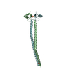


 PDBj
PDBj


 Assembly


 Mass: 96.063 Da / Num. of mol.: 2 / Source method: obtained synthetically / Formula: SO4
 Mass: 154.251 Da / Num. of mol.: 2 / Source method: obtained synthetically / Formula: C4H10O2S2
 Mass: 18.015 Da / Num. of mol.: 117 / Source method: isolated from a natural source / Formula: H2O
 Sample preparation
 Processing