 Movie
Movie Controller

[English] 日本語
 Yorodumi
- PDB-7aln: Cryo-EM structure of the divergent actomyosin complex from Plasmo... -
+ Open data

- Basic information
| Entry | Database: PDB / ID: 7aln | ||||||||||||
|---|---|---|---|---|---|---|---|---|---|---|---|---|---|
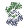
| Title | Cryo-EM structure of the divergent actomyosin complex from Plasmodium falciparum Myosin A in the Rigor state | ||||||||||||
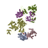
 Components Components |
| ||||||||||||
 Keywords Keywords | MOTOR PROTEIN / Malaria / Plasmodium falciparum / Myosin A / invasion / Actin 1 | ||||||||||||
| Function / homology |  Function and homology information Function and homology informationplastid inheritance / schizogony / pellicle / inner membrane pellicle complex / glideosome / symbiont-mediated actin polymerization-dependent cell-to-cell migration in host / HSP90 chaperone cycle for steroid hormone receptors (SHR) in the presence of ligand / Neutrophil degranulation / entry into host cell by a symbiont-containing vacuole / myosin complex ...plastid inheritance / schizogony / pellicle / inner membrane pellicle complex / glideosome / symbiont-mediated actin polymerization-dependent cell-to-cell migration in host / HSP90 chaperone cycle for steroid hormone receptors (SHR) in the presence of ligand / Neutrophil degranulation / entry into host cell by a symbiont-containing vacuole / myosin complex / microfilament motor activity / cytoskeletal motor activity / cytoskeleton organization / actin filament organization / actin filament / structural constituent of cytoskeleton / Hydrolases; Acting on acid anhydrides; Acting on acid anhydrides to facilitate cellular and subcellular movement / actin filament binding / actin cytoskeleton / actin binding / ATP hydrolysis activity / ATP binding / membrane / nucleus / plasma membrane / cytoplasm Similarity search - Function | ||||||||||||
| Biological species |  | ||||||||||||
| Method | ELECTRON MICROSCOPY / helical reconstruction / cryo EM / Resolution: 3.77 Å | ||||||||||||
 Authors Authors | Robert-Paganin, J. / Xu, X.-P. / Swift, M.F. / Auguin, D. / Robblee, J.P. / Lu, H. / Fagnant, P.M. / Krementsova, E.B. / Trybus, K.M. / Houdusse, A. ...Robert-Paganin, J. / Xu, X.-P. / Swift, M.F. / Auguin, D. / Robblee, J.P. / Lu, H. / Fagnant, P.M. / Krementsova, E.B. / Trybus, K.M. / Houdusse, A. / Volkmann, N. / Hanein, D. | ||||||||||||
| Funding support | 3items
| ||||||||||||
 Citation Citation | Journal: Nat Commun / Year: 2021 Title: The actomyosin interface contains an evolutionary conserved core and an ancillary interface involved in specificity. Authors: Julien Robert-Paganin / Xiao-Ping Xu / Mark F Swift / Daniel Auguin / James P Robblee / Hailong Lu / Patricia M Fagnant / Elena B Krementsova / Kathleen M Trybus / Anne Houdusse / Niels ...Authors: Julien Robert-Paganin / Xiao-Ping Xu / Mark F Swift / Daniel Auguin / James P Robblee / Hailong Lu / Patricia M Fagnant / Elena B Krementsova / Kathleen M Trybus / Anne Houdusse / Niels Volkmann / Dorit Hanein /   Abstract: Plasmodium falciparum, the causative agent of malaria, moves by an atypical process called gliding motility. Actomyosin interactions are central to gliding motility. However, the details of these ...Plasmodium falciparum, the causative agent of malaria, moves by an atypical process called gliding motility. Actomyosin interactions are central to gliding motility. However, the details of these interactions remained elusive until now. Here, we report an atomic structure of the divergent Plasmodium falciparum actomyosin system determined by electron cryomicroscopy at the end of the powerstroke (Rigor state). The structure provides insights into the detailed interactions that are required for the parasite to produce the force and motion required for infectivity. Remarkably, the footprint of the myosin motor on filamentous actin is conserved with respect to higher eukaryotes, despite important variability in the Plasmodium falciparum myosin and actin elements that make up the interface. Comparison with other actomyosin complexes reveals a conserved core interface common to all actomyosin complexes, with an ancillary interface involved in defining the spatial positioning of the motor on actin filaments. | ||||||||||||
| History |
|
- Structure visualization
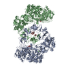
| Movie |
Movie viewer |
|---|---|
| Structure viewer | Molecule: MolmilJmol/JSmol |
- Downloads & links
-Download
| PDBx/mmCIF format | 7aln.cif.gz | 455.7 KB | Display | PDBx/mmCIF format |
|---|---|---|---|---|
| PDB format | pdb7aln.ent.gz | 376.2 KB | Display | PDB format |
| PDBx/mmJSON format | 7aln.json.gz | Tree view | PDBx/mmJSON format | |
| Others |  Other downloads Other downloads |
-Validation report
| Arichive directory | https://data.pdbj.org/pub/pdb/validation_reports/al/7alnftp://data.pdbj.org/pub/pdb/validation_reports/al/7aln | HTTPS FTP |
|---|
-Related structure data
| Related structure data | 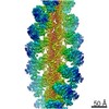 11818MC M: map data used to model this data C: citing same article ( |
|---|---|
| Similar structure data |


-Links
 PDBj
PDBj


- Assembly
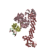
| Deposited unit | 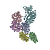
|
|---|---|
| 1 |
|
-Components
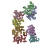
| #1: Protein | Mass: 41919.547 Da / Num. of mol.: 5 Source method: isolated from a genetically manipulated source Source: (gene. exp.) Strain: isolate 3D7 / Gene: PFL2215w / Production host:   Spodoptera frugiperda (fall armyworm) / References: UniProt: Q8I4X0 Spodoptera frugiperda (fall armyworm) / References: UniProt: Q8I4X0#2: Protein | | Mass: 92488.289 Da / Num. of mol.: 1 / Mutation: T417D Source method: isolated from a genetically manipulated source Source: (gene. exp.) Strain: isolate 3D7 / Gene: PF13_0233 / Production host: Spodoptera frugiperda (fall armyworm) / References: UniProt: Q8IDR3#3: Chemical | ChemComp-ADP /   Mass: 427.201 Da / Num. of mol.: 5 / Source method: obtained synthetically / Formula: C10H15N5O10P2 / Comment: ADP, energy-carrying molecule*YM Mass: 427.201 Da / Num. of mol.: 5 / Source method: obtained synthetically / Formula: C10H15N5O10P2 / Comment: ADP, energy-carrying molecule*YM#4: Chemical | ChemComp-MG /   Mass: 24.305 Da / Num. of mol.: 5 / Source method: obtained synthetically / Formula: Mg Mass: 24.305 Da / Num. of mol.: 5 / Source method: obtained synthetically / Formula: Mg#5: Chemical |   Mass: 709.670 Da / Num. of mol.: 3 / Source method: obtained synthetically / Formula: C36H45BrN4O6 Mass: 709.670 Da / Num. of mol.: 3 / Source method: obtained synthetically / Formula: C36H45BrN4O6Has ligand of interest | N | Has protein modification | Y | |
|---|
-Experimental details
-Experiment
| Experiment | Method: ELECTRON MICROSCOPY |
|---|---|
| EM experiment | Aggregation state: FILAMENT / 3D reconstruction method: helical reconstruction |
- Sample preparation
| Component | Name: actomyosin complex from Plasmodium falciparum / Type: COMPLEX / Entity ID: #1-#2 / Source: RECOMBINANT |
|---|---|
| Molecular weight | Experimental value: NO |
| Source (natural) | Organism: |
| Source (recombinant) | Organism: Spodoptera frugiperda (fall armyworm) |
| Buffer solution | pH: 7.4 |
| Specimen | Embedding applied: NO / Shadowing applied: NO / Staining applied: NO / Vitrification applied: YES |
| Vitrification | Cryogen name: ETHANE |
- Electron microscopy imaging
| Experimental equipment |  Model: Titan Krios / Image courtesy: FEI Company |
|---|---|
| Microscopy | Model: FEI TITAN KRIOS |
| Electron gun | Electron source:  FIELD EMISSION GUN / Accelerating voltage: 300 kV / Illumination mode: FLOOD BEAM FIELD EMISSION GUN / Accelerating voltage: 300 kV / Illumination mode: FLOOD BEAM |
| Electron lens | Mode: BRIGHT FIELD |
| Image recording | Electron dose: 60 e/Å2 / Film or detector model: FEI FALCON II (4k x 4k) |
- Processing
| CTF correction | Type: PHASE FLIPPING AND AMPLITUDE CORRECTION |
|---|---|
| Helical symmerty | Angular rotation/subunit: -168.1 ° / Axial rise/subunit: 27.3 Å / Axial symmetry: C1 |
| 3D reconstruction | Resolution: 3.77 Å / Resolution method: FSC 0.143 CUT-OFF / Num. of particles: 464646 / Symmetry type: HELICAL |
