 Movie
Movie Controller

[English] 日本語
 Yorodumi
- PDB-7al7: The Crystal Structure of Human IL-18 in Complex With Human IL-18 ... -
+ Open data

- Basic information
| Entry | Database: PDB / ID: 7al7 | ||||||
|---|---|---|---|---|---|---|---|
| Title | The Crystal Structure of Human IL-18 in Complex With Human IL-18 Binding Protein | ||||||
 Components Components |
| ||||||
 Keywords Keywords | CYTOKINE / decoy receptor / antagonist / complex | ||||||
| Function / homology |  Function and homology information Function and homology informationinterleukin-18 binding / interleukin-18 receptor binding / Interleukin-18 signaling / receptor antagonist activity / positive regulation of tissue remodeling / positive regulation of T-helper 1 cell cytokine production / positive regulation of T-helper 2 cell differentiation / positive regulation of interleukin-13 production / interleukin-18-mediated signaling pathway / positive regulation of neuroinflammatory response ...interleukin-18 binding / interleukin-18 receptor binding / Interleukin-18 signaling / receptor antagonist activity / positive regulation of tissue remodeling / positive regulation of T-helper 1 cell cytokine production / positive regulation of T-helper 2 cell differentiation / positive regulation of interleukin-13 production / interleukin-18-mediated signaling pathway / positive regulation of neuroinflammatory response / neutrophil activation / negative regulation of myoblast differentiation / Interleukin-1 processing / positive regulation of NK T cell proliferation / Interleukin-37 signaling / sleep / positive regulation of macrophage derived foam cell differentiation / natural killer cell activation / positive regulation of granulocyte macrophage colony-stimulating factor production / type 2 immune response / triglyceride homeostasis / T-helper 1 type immune response / positive regulation of tyrosine phosphorylation of STAT protein / natural killer cell mediated cytotoxicity / Interleukin-10 signaling / positive regulation of interleukin-17 production / positive regulation of activated T cell proliferation / positive regulation of natural killer cell proliferation / glutathione transferase / glutathione transferase activity / Pyroptosis / establishment of skin barrier / regulation of cell adhesion / Purinergic signaling in leishmaniasis infection / positive regulation of chemokine production / glutathione metabolic process / positive regulation of smooth muscle cell proliferation / cholesterol homeostasis / cytokine activity / positive regulation of non-canonical NF-kappaB signal transduction / positive regulation of NF-kappaB transcription factor activity / positive regulation of type II interferon production / cellular response to hydrogen peroxide / cytokine-mediated signaling pathway / positive regulation of inflammatory response / cellular response to tumor necrosis factor / cell-cell signaling / positive regulation of cold-induced thermogenesis / cellular response to lipopolysaccharide / angiogenesis / Interleukin-4 and Interleukin-13 signaling / response to lipopolysaccharide / cell population proliferation / positive regulation of phosphatidylinositol 3-kinase/protein kinase B signal transduction / defense response to Gram-positive bacterium / immune response / inflammatory response / positive regulation of transcription by RNA polymerase II / extracellular space / extracellular exosome / extracellular region / cytosol Similarity search - Function | ||||||
| Biological species |  Homo sapiens (human) Homo sapiens (human) | ||||||
| Method |  X-RAY DIFFRACTION / SYNCHROTRON / MOLECULAR REPLACEMENT / Resolution: 1.801 Å X-RAY DIFFRACTION / SYNCHROTRON / MOLECULAR REPLACEMENT / Resolution: 1.801 Å | ||||||
 Authors Authors | Detry, S. / Andries, J. / Bloch, Y. / Clancy, D. / Savvides, S.N. | ||||||
| Funding support | European Union, 1items
| ||||||
 Citation Citation | Journal: J.Biol.Chem. / Year: 2022 Title: Structural basis of human IL-18 sequestration by the decoy receptor IL-18 binding protein in inflammation and tumor immunity. Authors: Detry, S. / Andries, J. / Bloch, Y. / Gabay, C. / Clancy, D.M. / Savvides, S.N. | ||||||
| History |
|
- Structure visualization
| Structure viewer | Molecule: MolmilJmol/JSmol |
|---|
- Downloads & links
-Download
| PDBx/mmCIF format | 7al7.cif.gz | 179.7 KB | Display | PDBx/mmCIF format |
|---|---|---|---|---|
| PDB format | pdb7al7.ent.gz | 139.4 KB | Display | PDB format |
| PDBx/mmJSON format | 7al7.json.gz | Tree view | PDBx/mmJSON format | |
| Others |  Other downloads Other downloads |
-Validation report
| Summary document | 7al7_validation.pdf.gz | 463.6 KB | Display | wwPDB validaton report |
|---|---|---|---|---|
| Full document | 7al7_full_validation.pdf.gz | 464.8 KB | Display | |
| Data in XML | 7al7_validation.xml.gz | 13.4 KB | Display | |
| Data in CIF | 7al7_validation.cif.gz | 18.7 KB | Display | |
| Arichive directory | https://data.pdbj.org/pub/pdb/validation_reports/al/7al7ftp://data.pdbj.org/pub/pdb/validation_reports/al/7al7 | HTTPS FTP |
-Related structure data
| Related structure data |  3f62S S: Starting model for refinement |
|---|---|
| Similar structure data |
-Links
 PDBj
PDBj


- Assembly
| Deposited unit | 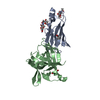
| ||||||||
|---|---|---|---|---|---|---|---|---|---|
| 1 |
| ||||||||
| Unit cell |
|
-Components
| #1: Protein | Mass: 18749.832 Da / Num. of mol.: 1 Source method: isolated from a genetically manipulated source Details: The final protein sequence used for crystallization starts at T30 and ends at D170. The N-terminal RTPmu signal sequence is cleaved during expression. The C-terminal AVI-HIS tag was cleaved ...Details: The final protein sequence used for crystallization starts at T30 and ends at D170. The N-terminal RTPmu signal sequence is cleaved during expression. The C-terminal AVI-HIS tag was cleaved by caspase-3 after the DEVD site. In reference to the native sequence, the final protein sequence starts at Q63 and ends at G194, as it lacks an N-terminal region from T31 to K62. Source: (gene. exp.) Homo sapiens (human) / Gene: IL18BP / Plasmid: pHLSec / Cell line (production host): HEK293S MGAT -/- / Production host: Homo sapiens (human) / References: UniProt: O95998 | ||||||
|---|---|---|---|---|---|---|---|
| #2: Protein | Mass: 45695.297 Da / Num. of mol.: 1 Source method: isolated from a genetically manipulated source Details: The protein was expressed as a HIS-GST fusion which was cleaved after the DEVD site using caspasea-3. Thus, the final protein sequence used for crystallization starts at Y234 and ends at ...Details: The protein was expressed as a HIS-GST fusion which was cleaved after the DEVD site using caspasea-3. Thus, the final protein sequence used for crystallization starts at Y234 and ends at D390. In reference to the native sequence, the final protein sequence starts at Y37 and ends at D193,The protein was expressed as a HIS-GST fusion which was cleaved after the DEVD site using caspasea-3. Thus, the final protein sequence used for crystallization starts at Y234 and ends at D390. In reference to the native sequence, the final protein sequence starts at Y37 and ends at D193,The protein was expressed as a HIS-GST fusion which was cleaved after the DEVD site using caspasea-3. Thus, the final protein sequence used for crystallization starts at Y234 and ends at D390. In reference to the native sequence, the final protein sequence starts at Y37 and ends at D193,The protein was expressed as a HIS-GST fusion which was cleaved after the DEVD site using caspasea-3. Thus, the final protein sequence used for crystallization starts at Y234 and ends at D390. In reference to the native sequence, the final protein sequence starts at Y37 and ends at D193,The protein was expressed as a HIS-GST fusion which was cleaved after the DEVD site using caspasea-3. Thus, the final protein sequence used for crystallization starts at Y234 and ends at D390. In reference to the native sequence, the final protein sequence starts at Y37 and ends at D193,The protein was expressed as a HIS-GST fusion which was cleaved after the DEVD site using caspasea-3. Thus, the final protein sequence used for crystallization starts at Y234 and ends at D390. In reference to the native sequence, the final protein sequence starts at Y37 and ends at D193,The protein was expressed as a HIS-GST fusion which was cleaved after the DEVD site using caspasea-3. Thus, the final protein sequence used for crystallization starts at Y234 and ends at D390. In reference to the native sequence, the final protein sequence starts at Y37 and ends at D193,The protein was expressed as a HIS-GST fusion which was cleaved after the DEVD site using caspasea-3. Thus, the final protein sequence used for crystallization starts at Y234 and ends at D390. In reference to the native sequence, the final protein sequence starts at Y37 and ends at D193 Source: (gene. exp.) Homo sapiens (human)Plasmid: pET-42 / Gene: IL18, IGIF, IL1F4 / Details (production host): HIS-GST tag added N-terminally / Production host:  References: UniProt: P08515, UniProt: Q14116, glutathione transferase | ||||||
| #3: Sugar |   Type: D-saccharide, beta linking / Mass: 221.208 Da / Num. of mol.: 3 / Source method: obtained synthetically / Formula: C8H15NO6 Type: D-saccharide, beta linking / Mass: 221.208 Da / Num. of mol.: 3 / Source method: obtained synthetically / Formula: C8H15NO6#4: Water | ChemComp-HOH / |  Mass: 18.015 Da / Num. of mol.: 130 / Source method: isolated from a natural source / Formula: H2O Mass: 18.015 Da / Num. of mol.: 130 / Source method: isolated from a natural source / Formula: H2OHas ligand of interest | N | Has protein modification | Y | |
-Experimental details
-Experiment
| Experiment | Method: X-RAY DIFFRACTION / Number of used crystals: 1 |
|---|
- Sample preparation
| Crystal grow | Temperature: 293 K / Method: vapor diffusion / pH: 6.5 Details: 0.2 M Calcium acetate hydrate, 0.1 M Sodium cacodylate trihydrate pH 6.5, 18% w/v Polyethylene glycol 8000 Temp details: Room temperature |
|---|
-Data collection
| Diffraction | Mean temperature: 100 K / Serial crystal experiment: N | ||||||||||||||||||||||||||||||||||||||||||||||||||||||||||||||||||||||
|---|---|---|---|---|---|---|---|---|---|---|---|---|---|---|---|---|---|---|---|---|---|---|---|---|---|---|---|---|---|---|---|---|---|---|---|---|---|---|---|---|---|---|---|---|---|---|---|---|---|---|---|---|---|---|---|---|---|---|---|---|---|---|---|---|---|---|---|---|---|---|---|
| Diffraction source | Source: SYNCHROTRON / Site: PETRA III, EMBL c/o DESY  / Beamline: P14 (MX2) / Wavelength: 0.9762 Å / Beamline: P14 (MX2) / Wavelength: 0.9762 Å | ||||||||||||||||||||||||||||||||||||||||||||||||||||||||||||||||||||||
| Detector | Type: DECTRIS EIGER X 16M / Detector: PIXEL / Date: May 24, 2019 | ||||||||||||||||||||||||||||||||||||||||||||||||||||||||||||||||||||||
| Radiation | Protocol: SINGLE WAVELENGTH / Monochromatic (M) / Laue (L): M / Scattering type: x-ray | ||||||||||||||||||||||||||||||||||||||||||||||||||||||||||||||||||||||
| Radiation wavelength | Wavelength: 0.9762 Å / Relative weight: 1 | ||||||||||||||||||||||||||||||||||||||||||||||||||||||||||||||||||||||
| Reflection | Resolution: 1.8→59.39 Å / Num. obs: 26507 / % possible obs: 98.27 % / Redundancy: 6.9 % / CC1/2: 0.999 / CC star: 1 / Rmerge(I) obs: 0.048 / Rpim(I) all: 0.02 / Rrim(I) all: 0.053 / Net I/σ(I): 15.67 | ||||||||||||||||||||||||||||||||||||||||||||||||||||||||||||||||||||||
| Reflection shell |
|
- Processing
| Software |
| ||||||||||||||||||||||||||||||||||||||||||||||||||||||||||||||||||||||||||||||||||||||||||||||||||||||||||||||||||||||||
|---|---|---|---|---|---|---|---|---|---|---|---|---|---|---|---|---|---|---|---|---|---|---|---|---|---|---|---|---|---|---|---|---|---|---|---|---|---|---|---|---|---|---|---|---|---|---|---|---|---|---|---|---|---|---|---|---|---|---|---|---|---|---|---|---|---|---|---|---|---|---|---|---|---|---|---|---|---|---|---|---|---|---|---|---|---|---|---|---|---|---|---|---|---|---|---|---|---|---|---|---|---|---|---|---|---|---|---|---|---|---|---|---|---|---|---|---|---|---|---|---|---|
| Refinement | Method to determine structure: MOLECULAR REPLACEMENT Starting model: 3F62 Resolution: 1.801→59.386 Å / SU ML: 0.29 / Cross valid method: THROUGHOUT / σ(F): 1.36 / Phase error: 39.86 / Stereochemistry target values: ML
| ||||||||||||||||||||||||||||||||||||||||||||||||||||||||||||||||||||||||||||||||||||||||||||||||||||||||||||||||||||||||
| Solvent computation | Shrinkage radii: 0.9 Å / VDW probe radii: 1.11 Å / Solvent model: FLAT BULK SOLVENT MODEL | ||||||||||||||||||||||||||||||||||||||||||||||||||||||||||||||||||||||||||||||||||||||||||||||||||||||||||||||||||||||||
| Displacement parameters | Biso max: 184.21 Å2 / Biso mean: 38.4789 Å2 / Biso min: 5.01 Å2 | ||||||||||||||||||||||||||||||||||||||||||||||||||||||||||||||||||||||||||||||||||||||||||||||||||||||||||||||||||||||||
| Refinement step | Cycle: final / Resolution: 1.801→59.386 Å
| ||||||||||||||||||||||||||||||||||||||||||||||||||||||||||||||||||||||||||||||||||||||||||||||||||||||||||||||||||||||||
| LS refinement shell | Refine-ID: X-RAY DIFFRACTION / Rfactor Rfree error: 0
|
