[English] 日本語
Yorodumi
- PDB-7ajp: Crystal Structure of Human Adenovirus 56 Fiber Knob -

+
Open data


ID or keywords:

Loading...

-
Basic information

Entry
Database: PDB / ID: 7ajp
TitleCrystal Structure of Human Adenovirus 56 Fiber Knob
ComponentsFiber
KeywordsVIRAL PROTEIN / Human Adenovirus / Fiber Knob
Function / homology
Function and homology information


viral capsid / cell adhesion / symbiont entry into host cell / host cell nucleus / virion attachment to host cell
Similarity search - Function
Adenoviral fibre protein, knob / Adenoviral fibre protein (knob domain) / Adenovirus fibre protein / Attachment protein shaft domain superfamily / Adenovirus pIV-like, attachment domain
Similarity search - Domain/homology
NITRATE ION / TRIETHYLENE GLYCOL / Fiber
Similarity search - Component
Biological speciesHuman adenovirus 56
MethodX-RAY DIFFRACTION / SYNCHROTRON / MOLECULAR REPLACEMENT / Resolution: 1.38201802428 Å
AuthorsStrebl, M. / Mindler, K. / Stehle, T.
Funding support Germany, 1items
OrganizationGrant numberCountry
Baden-Wuerttemberg-Stiftung Germany
Citation
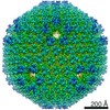
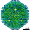
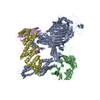
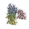
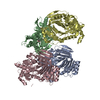
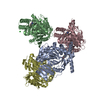
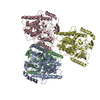
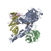
Journal: Proc Natl Acad Sci U S A / Year: 2021
Title: Human species D adenovirus hexon capsid protein mediates cell entry through a direct interaction with CD46.
Authors: B David Persson / Lijo John / Karim Rafie / Michael Strebl / Lars Frängsmyr / Monika Z Ballmann / Katja Mindler / Menzo Havenga / Angelique Lemckert / Thilo Stehle / Lars-Anders Carlson / Niklas Arnberg /
Abstract: Human adenovirus species D (HAdV-D) types are currently being explored as vaccine vectors for coronavirus disease 2019 (COVID-19) and other severe infectious diseases. The efficacy of such vector- ...Human adenovirus species D (HAdV-D) types are currently being explored as vaccine vectors for coronavirus disease 2019 (COVID-19) and other severe infectious diseases. The efficacy of such vector-based vaccines depends on functional interactions with receptors on host cells. Adenoviruses of different species are assumed to enter host cells mainly by interactions between the knob domain of the protruding fiber capsid protein and cellular receptors. Using a cell-based receptor-screening assay, we identified CD46 as a receptor for HAdV-D56. The function of CD46 was validated in infection experiments using cells lacking and overexpressing CD46, and by competition infection experiments using soluble CD46. Remarkably, unlike HAdV-B types that engage CD46 through interactions with the knob domain of the fiber protein, HAdV-D types infect host cells through a direct interaction between CD46 and the hexon protein. Soluble hexon proteins (but not fiber knob) inhibited HAdV-D56 infection, and surface plasmon analyses demonstrated that CD46 binds to HAdV-D hexon (but not fiber knob) proteins. Cryoelectron microscopy analysis of the HAdV-D56 virion-CD46 complex confirmed the interaction and showed that CD46 binds to the central cavity of hexon trimers. Finally, soluble CD46 inhibited infection by 16 out of 17 investigated HAdV-D types, suggesting that CD46 is an important receptor for a large group of adenoviruses. In conclusion, this study identifies a noncanonical entry mechanism used by human adenoviruses, which adds to the knowledge of adenovirus biology and can also be useful for development of adenovirus-based vaccine vectors.
History
DepositionSep 29, 2020Deposition site: PDBE / Processing site: PDBE
Revision 1.0Jan 13, 2021Provider: repository / Type: Initial release
Revision 1.1Jan 31, 2024Group: Data collection / Database references / Refinement description
Category: chem_comp_atom / chem_comp_bond ...chem_comp_atom / chem_comp_bond / database_2 / pdbx_initial_refinement_model
Item: _database_2.pdbx_DOI / _database_2.pdbx_database_accession

-
Structure visualization

Structure viewerMolecule:
MolmilJmol/JSmol

Downloads & links

-
Assembly

Deposited unit
A: Fiber
B: Fiber
C: Fiber
D: Fiber
E: Fiber
F: Fiber
G: Fiber
H: Fiber
I: Fiber
J: Fiber
K: Fiber
L: Fiber
hetero molecules


Theoretical massNumber of molelcules
Total (without water)285,90873
Polymers281,14112
Non-polymers4,76761
Water38,7862153
1


  • Idetical with deposited unit
  • defined by author
  • Evidence: gel filtration, light scattering
TypeNameSymmetry operationNumber
identity operation1_555x,y,z1
Unit cell
Length a, b, c (Å)132.443, 182.529, 117.029
Angle α, β, γ (deg.)90.000, 118.541, 90.000
Int Tables number5
Space group name H-MC121
Space group name HallC2y
Symmetry operation#1: x,y,z
#2: -x,y,-z
#3: x+1/2,y+1/2,z
#4: -x+1/2,y+1/2,-z

-
Components

-
Protein , 1 types, 12 molecules ABCDEFGHIJKL

#1: Protein
Fiber /


Mass: 23428.441 Da / Num. of mol.: 12
Source method: isolated from a genetically manipulated source
Source: (gene. exp.) Human adenovirus 56 / Production host: Escherichia coli (E. coli) / References: UniProt: R9RU05

-
Non-polymers , 5 types, 2214 molecules

#2: Chemical
ChemComp-GOL / GLYCEROL / GLYCERIN / PROPANE-1,2,3-TRIOL / Glycerol


Mass: 92.094 Da / Num. of mol.: 18 / Source method: obtained synthetically / Formula: C3H8O3
#3: Chemical
ChemComp-NO3 / NITRATE ION / Nitrate


Mass: 62.005 Da / Num. of mol.: 5 / Source method: obtained synthetically / Formula: NO3
#4: Chemical...
ChemComp-EDO / 1,2-ETHANEDIOL / ETHYLENE GLYCOL / Ethylene glycol


Mass: 62.068 Da / Num. of mol.: 33 / Source method: obtained synthetically / Formula: C2H6O2
#5: Chemical
ChemComp-PGE / TRIETHYLENE GLYCOL / Polyethylene glycol


Mass: 150.173 Da / Num. of mol.: 5 / Source method: obtained synthetically / Formula: C6H14O4
#6: Water ChemComp-HOH / water / Water


Mass: 18.015 Da / Num. of mol.: 2153 / Source method: isolated from a natural source / Formula: H2O

-
Details

Has ligand of interestN

-
Experimental details

-
Experiment

ExperimentMethod: X-RAY DIFFRACTION / Number of used crystals: 1

-
Sample preparation

CrystalDensity Matthews: 2.41 Å3/Da / Density % sol: 48.96 %
Crystal growTemperature: 277 K / Method: vapor diffusion, sitting drop / pH: 7.4 / Details: 20 % PEG3350, 200 mM magnesium nitrate hexahydrate

-
Data collection

DiffractionMean temperature: 100 K / Serial crystal experiment: N
Diffraction sourceSource: SYNCHROTRON / Site: SLS / Beamline: X06DA / Wavelength: 1 Å
DetectorType: DECTRIS PILATUS 2M / Detector: PIXEL / Date: Oct 6, 2018
RadiationProtocol: SINGLE WAVELENGTH / Monochromatic (M) / Laue (L): M / Scattering type: x-ray
Radiation wavelengthWavelength: 1 Å / Relative weight: 1
ReflectionResolution: 1.38→49.1 Å / Num. obs: 491039 / % possible obs: 98.9 % / Redundancy: 6.8 % / Biso Wilson estimate: 18.4589513852 Å2 / CC1/2: 0.999 / Rrim(I) all: 0.086 / Net I/σ(I): 12.41
Reflection shellResolution: 1.38→1.46 Å / Redundancy: 6.5 % / Mean I/σ(I) obs: 1.15 / Num. unique obs: 80171 / CC1/2: 0.479 / Rrim(I) all: 1.461 / % possible all: 97

-
Processing

Software
NameVersionClassification
PHENIX1.10.1_2155refinement
XDSdata reduction
XDSdata scaling
PHASERphasing
RefinementMethod to determine structure: MOLECULAR REPLACEMENT
Starting model: 1UXE
Resolution: 1.38201802428→42.6447882419 Å / SU ML: 0.180530063572 / Cross valid method: FREE R-VALUE / σ(F): 1.33837135366 / Phase error: 16.5201204278
Stereochemistry target values: GeoStd + Monomer Library + CDL v1.2
RfactorNum. reflection% reflection
Rfree0.166873226508 7365 1.49998472521 %
Rwork0.134150253001 483640 -
obs0.134641935546 491005 99.0276830985 %
Solvent computationShrinkage radii: 0.9 Å / VDW probe radii: 1.11 Å / Solvent model: FLAT BULK SOLVENT MODEL
Displacement parametersBiso mean: 27.7005491595 Å2
Refinement stepCycle: LAST / Resolution: 1.38201802428→42.6447882419 Å
ProteinNucleic acidLigandSolventTotal
Num. atoms17316 0 292 2153 19761
Refine LS restraints
Refine-IDTypeDev idealNumber
X-RAY DIFFRACTIONf_bond_d0.010020833170818494
X-RAY DIFFRACTIONf_angle_d1.086232568525157
X-RAY DIFFRACTIONf_chiral_restr0.09670725140172847
X-RAY DIFFRACTIONf_plane_restr0.006713435001553193
X-RAY DIFFRACTIONf_dihedral_angle_d14.90075520896730
LS refinement shell
Resolution (Å)Rfactor RfreeNum. reflection RfreeRfactor RworkNum. reflection RworkRefine-ID% reflection obs (%)
1.38201802428-1.39770.3578766577212300.32280701182115095X-RAY DIFFRACTION92.9576610457
1.3977-1.41420.3442123598932430.31394860431215933X-RAY DIFFRACTION98.0482482725
1.4142-1.43140.3107754836322420.29593264155215918X-RAY DIFFRACTION98.1893304168
1.4314-1.44950.2779336628362440.26204919147615988X-RAY DIFFRACTION98.1972171809
1.4495-1.46860.2986738186132430.23895841735916013X-RAY DIFFRACTION98.2591876209
1.4686-1.48870.2658281795672430.22340805212315956X-RAY DIFFRACTION98.3725025809
1.4887-1.510.2661483255622450.20257466215116037X-RAY DIFFRACTION98.6070736434
1.51-1.53250.2435349202262440.18803156877516024X-RAY DIFFRACTION98.6836518047
1.5325-1.55650.2053337173412440.17365827943616049X-RAY DIFFRACTION98.6378496186
1.5565-1.5820.2075794423342460.16918757604316119X-RAY DIFFRACTION98.8105301292
1.582-1.60930.2077986422172440.16211781137716022X-RAY DIFFRACTION98.9596641723
1.6093-1.63860.2008977802792450.15507325164116109X-RAY DIFFRACTION98.9412547644
1.6386-1.67010.1872245971832450.15220975058416089X-RAY DIFFRACTION99.1381403253
1.6701-1.70420.1903336858932460.143688819716165X-RAY DIFFRACTION99.1122116198
1.7042-1.74120.1699868796012450.12878862790616110X-RAY DIFFRACTION99.2897037397
1.7412-1.78170.1673294367712460.12552601164116160X-RAY DIFFRACTION99.3881383655
1.7817-1.82630.1476515870292470.11387337307216195X-RAY DIFFRACTION99.4736523686
1.8263-1.87570.1557192106352460.11232707907216182X-RAY DIFFRACTION99.5214151572
1.8757-1.93080.1549897216872470.10875353567516181X-RAY DIFFRACTION99.6240145543
1.9308-1.99320.1472123846882470.10993674609416233X-RAY DIFFRACTION99.6492925384
1.9932-2.06440.1457751046792480.11268734852816298X-RAY DIFFRACTION99.7528184723
2.0644-2.14710.1536254388772470.11394160609216195X-RAY DIFFRACTION99.8481812109
2.1471-2.24480.1567143320012480.11463092932616285X-RAY DIFFRACTION99.903317421
2.2448-2.36310.1475578645342480.12147725158916277X-RAY DIFFRACTION99.9213931552
2.3631-2.51110.1746331879942480.1258732113316324X-RAY DIFFRACTION99.945721006
2.5111-2.7050.1488742488772480.12973369048916269X-RAY DIFFRACTION99.8730197122
2.705-2.97720.1657203400472480.13350314586316295X-RAY DIFFRACTION99.9818687296
2.9772-3.40780.1517503397042490.12973262322716328X-RAY DIFFRACTION99.9638183682
3.4078-4.29280.1516023577282490.12085572482616350X-RAY DIFFRACTION99.9518275426
4.2928-42.64478824190.1534935300382500.13097915361716441X-RAY DIFFRACTION99.790744948

+
About Yorodumi

-
News

-
Feb 9, 2022. New format data for meta-information of EMDB entries

New format data for meta-information of EMDB entries

  • Version 3 of the EMDB header file is now the official format.
  • The previous official version 1.9 will be removed from the archive.

Related info.:EMDB header

External links:wwPDB to switch to version 3 of the EMDB data model

-
Aug 12, 2020. Covid-19 info

Covid-19 info

URL: https://pdbj.org/emnavi/covid19.php

New page: Covid-19 featured information page in EM Navigator.

Related info.:Covid-19 info / Mar 5, 2020. Novel coronavirus structure data

+
Mar 5, 2020. Novel coronavirus structure data

Novel coronavirus structure data

Related info.:Yorodumi Speices / Aug 12, 2020. Covid-19 info

External links:COVID-19 featured content - PDBj / Molecule of the Month (242):Coronavirus Proteases

+
Jan 31, 2019. EMDB accession codes are about to change! (news from PDBe EMDB page)

EMDB accession codes are about to change! (news from PDBe EMDB page)

  • The allocation of 4 digits for EMDB accession codes will soon come to an end. Whilst these codes will remain in use, new EMDB accession codes will include an additional digit and will expand incrementally as the available range of codes is exhausted. The current 4-digit format prefixed with “EMD-” (i.e. EMD-XXXX) will advance to a 5-digit format (i.e. EMD-XXXXX), and so on. It is currently estimated that the 4-digit codes will be depleted around Spring 2019, at which point the 5-digit format will come into force.
  • The EM Navigator/Yorodumi systems omit the EMD- prefix.

Related info.:Q: What is EMD? / ID/Accession-code notation in Yorodumi/EM Navigator

External links:EMDB Accession Codes are Changing Soon! / Contact to PDBj

+
Jul 12, 2017. Major update of PDB

Major update of PDB

  • wwPDB released updated PDB data conforming to the new PDBx/mmCIF dictionary.
  • This is a major update changing the version number from 4 to 5, and with Remediation, in which all the entries are updated.
  • In this update, many items about electron microscopy experimental information are reorganized (e.g. em_software).
  • Now, EM Navigator and Yorodumi are based on the updated data.

External links:wwPDB Remediation / Enriched Model Files Conforming to OneDep Data Standards Now Available in the PDB FTP Archive

-
Yorodumi

Thousand views of thousand structures

  • Yorodumi is a browser for structure data from EMDB, PDB, SASBDB, etc.
  • This page is also the successor to EM Navigator detail page, and also detail information page/front-end page for Omokage search.
  • The word "yorodu" (or yorozu) is an old Japanese word meaning "ten thousand". "mi" (miru) is to see.

Related info.:EMDB / PDB / SASBDB / Comparison of 3 databanks / Yorodumi Search / Aug 31, 2016. New EM Navigator & Yorodumi / Yorodumi Papers / Jmol/JSmol / Function and homology information / Changes in new EM Navigator and Yorodumi

Read more