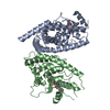
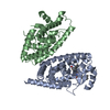
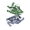
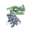
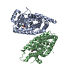
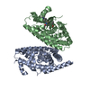
Entry Database : PDB / ID : 7ahjTitle Crystal structure of PPARgamma V290M mutant ligand binding domain in complex with farglitazar Peroxisome proliferator-activated receptor gamma Keywords / Function / homology Function Domain/homology Component
/ / / / / / / / / / / / / / / / / / / / / / / / / / / / / / / / / / / / / / / / / / / / / / / / / / / / / / / / / / / / / / / / / / / / / / / / / / / / / / / / / / / / / / / / / / / / / / / / / / / / / / / / / / / / / / / / / / / / / / / / / / / / / / / / / / / / / Biological species Homo sapiens (human)Method / / / / Resolution : 2.1 Å Authors Schoenmakers, E. / Schwabe, B.T.W. / Fairall, L. / Chatterjee, K. / Schwabe, J.W.R. Journal : To Be Published Title : Crystal structure of PPARgamma V290M mutant ligand binding domain in complex with farglitazarAuthors : Schoenmakers, E. / Schwabe, B.T.W. / Fairall, L. / Chatterjee, K. / Schwabe, J.W.R. History Deposition Sep 24, 2020 Deposition site / Processing site Revision 1.0 Oct 14, 2020 Provider / Type Revision 1.1 Jan 31, 2024 Group / Database references / Refinement descriptionCategory chem_comp_atom / chem_comp_bond ... chem_comp_atom / chem_comp_bond / database_2 / pdbx_initial_refinement_model Item / _database_2.pdbx_database_accession
Show all Show less
 Movie
Movie Controller

 Yorodumi
 Open data
 Basic information
 Components
 Keywords
 Function and homology information
 Homo sapiens (human)
 X-RAY DIFFRACTION /
  Authors
 Citation
 Structure visualization
 Downloads & links
 Other downloads
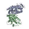


 PDBj
PDBj Assembly
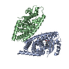


 Mass: 546.612 Da / Num. of mol.: 1 / Source method: obtained synthetically / Formula: C34H30N2O5
 Mass: 18.015 Da / Num. of mol.: 159 / Source method: isolated from a natural source / Formula: H2O
 Sample preparation
 / Beamline: ID23-2 / Wavelength: 0.8726 Å
 Processing