 Movie
Movie Controller


+ Open data

- Basic information
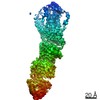
| Entry | Database: PDB / ID: 7ag9 | ||||||
|---|---|---|---|---|---|---|---|
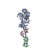
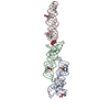
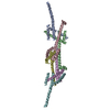
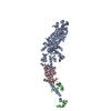
| Title | Structure of the Kar9 protein | ||||||
 Components Components | Kar9 | ||||||
 Keywords Keywords | CELL CYCLE / Cytoskeleton / Cell Division / Microtubules | ||||||
| Function / homology | Karyogamy protein, KAR9 / Yeast cortical protein KAR9 / Karyogamy protein KAR9 Function and homology information Function and homology information | ||||||
| Biological species |  Naumovozyma castellii (fungus) Naumovozyma castellii (fungus) | ||||||
| Method |  X-RAY DIFFRACTION / SYNCHROTRON / SAD / Resolution: 3.3 Å X-RAY DIFFRACTION / SYNCHROTRON / SAD / Resolution: 3.3 Å | ||||||
 Authors Authors | Kumar, A. / prota, A.E. / Steinmetz, M.O. | ||||||
| Funding support |  Switzerland, 1items Switzerland, 1items
| ||||||
 Citation Citation | Journal: Structure / Year: 2021 Title: Structure of the Kar9 protein Authors: Kumar, A. / Steinmetz, M.O. | ||||||
| History |
|
- Structure visualization
| Structure viewer | Molecule: MolmilJmol/JSmol |
|---|
- Downloads & links
-Download
| PDBx/mmCIF format | 7ag9.cif.gz | 160.2 KB | Display | PDBx/mmCIF format |
|---|---|---|---|---|
| PDB format | pdb7ag9.ent.gz | 125.1 KB | Display | PDB format |
| PDBx/mmJSON format | 7ag9.json.gz | Tree view | PDBx/mmJSON format | |
| Others |  Other downloads Other downloads |
-Validation report
| Summary document | 7ag9_validation.pdf.gz | 320.9 KB | Display | wwPDB validaton report |
|---|---|---|---|---|
| Full document | 7ag9_full_validation.pdf.gz | 331.9 KB | Display | |
| Data in XML | 7ag9_validation.xml.gz | 27 KB | Display | |
| Data in CIF | 7ag9_validation.cif.gz | 36 KB | Display | |
| Arichive directory | https://data.pdbj.org/pub/pdb/validation_reports/ag/7ag9ftp://data.pdbj.org/pub/pdb/validation_reports/ag/7ag9 | HTTPS FTP |
-Related structure data
| Similar structure data |
|---|

-Links
 PDBj
PDBj
- Assembly
| Deposited unit | 
| ||||||||
|---|---|---|---|---|---|---|---|---|---|
| 1 |
| ||||||||
| Unit cell |
|
-Components
| #1: Protein | Mass: 50291.051 Da / Num. of mol.: 2 Source method: isolated from a genetically manipulated source Source: (gene. exp.) Naumovozyma castellii (strain ATCC 76901 / CBS 4309 / NBRC 1992 / NRRL Y-12630) (fungus)Strain: ATCC 76901 / CBS 4309 / NBRC 1992 / NRRL Y-12630 / Gene: NCAS0D02220, NCAS_0D02220 / Production host:  |
|---|
-Experimental details
-Experiment
| Experiment | Method: X-RAY DIFFRACTION / Number of used crystals: 1 |
|---|
- Sample preparation
| Crystal | Density Matthews: 6.48 Å3/Da / Density % sol: 81.03 % |
|---|---|
| Crystal grow | Temperature: 293 K / Method: vapor diffusion, sitting drop Details: 20 mM Tris-HCl, pH 7.5, 500 mM NaCl supplemented with 250 mM gamma amino butyric acid (GABA). |
-Data collection
| Diffraction | Mean temperature: 100 K / Serial crystal experiment: N |
|---|---|
| Diffraction source | Source: SYNCHROTRON / Site: SLS / Beamline: X06DA / Wavelength: 1 Å |
| Detector | Type: DECTRIS PILATUS 2M / Detector: PIXEL / Date: Jun 29, 2016 |
| Radiation | Protocol: SINGLE WAVELENGTH / Monochromatic (M) / Laue (L): M / Scattering type: x-ray |
| Radiation wavelength | Wavelength: 1 Å / Relative weight: 1 |
| Reflection | Resolution: 3.3→50.01 Å / Num. obs: 38506 / % possible obs: 99.8 % / Redundancy: 12.3 % / CC1/2: 0.999 / Net I/σ(I): 11.09 |
| Reflection shell | Resolution: 3.3→3.4 Å / Num. unique obs: 12118 / CC1/2: 0.51 |
- Processing
| Software |
| ||||||||||||||||||||||||||||||||||||||||||||||||||||||||||||
|---|---|---|---|---|---|---|---|---|---|---|---|---|---|---|---|---|---|---|---|---|---|---|---|---|---|---|---|---|---|---|---|---|---|---|---|---|---|---|---|---|---|---|---|---|---|---|---|---|---|---|---|---|---|---|---|---|---|---|---|---|---|
| Refinement | Method to determine structure: SAD / Resolution: 3.3→46.86 Å / Cor.coef. Fo:Fc: 0.942 / Cor.coef. Fo:Fc free: 0.926 / SU B: 28.741 / SU ML: 0.442 / Cross valid method: THROUGHOUT / σ(F): 0 / ESU R: 0.625 / ESU R Free: 0.397 / Stereochemistry target values: MAXIMUM LIKELIHOOD Details: HYDROGENS HAVE BEEN ADDED IN THE RIDING POSITIONS U VALUES : REFINED INDIVIDUALLY
| ||||||||||||||||||||||||||||||||||||||||||||||||||||||||||||
| Solvent computation | Ion probe radii: 0.8 Å / Shrinkage radii: 0.8 Å / VDW probe radii: 1.2 Å / Solvent model: MASK | ||||||||||||||||||||||||||||||||||||||||||||||||||||||||||||
| Displacement parameters | Biso max: 306.78 Å2 / Biso mean: 144.18 Å2 / Biso min: 83.68 Å2
| ||||||||||||||||||||||||||||||||||||||||||||||||||||||||||||
| Refinement step | Cycle: final / Resolution: 3.3→46.86 Å
| ||||||||||||||||||||||||||||||||||||||||||||||||||||||||||||
| Refine LS restraints |
| ||||||||||||||||||||||||||||||||||||||||||||||||||||||||||||
| LS refinement shell | Resolution: 3.301→3.386 Å / Rfactor Rfree error: 0 / Total num. of bins used: 20
|
