 Movie
Movie Controller

[English] 日本語
 Yorodumi
- PDB-7ag5: Structure of the Laspartomycin C double mutant G4D D-allo-Thr9D-D... -
+ Open data

- Basic information
| Entry | Database: PDB / ID: 7ag5 | ||||||
|---|---|---|---|---|---|---|---|
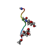
| Title | Structure of the Laspartomycin C double mutant G4D D-allo-Thr9D-Dap in complex with Geranyl phosphate | ||||||
 Components Components | Laspartomycin C double mutant G4D D-allo-Thr9D-Dap | ||||||
 Keywords Keywords | ANTIBIOTIC / Calcium-dependent antibiotic / Bacterial cell wall biosynthesis inhibitor / Lipopeptide antibiotic | ||||||
| Function / homology | (~{E})-13-methyltetradec-2-enoic acid / Geranyl phosphate Function and homology information Function and homology information | ||||||
| Biological species |  Streptomyces viridochromogenes (bacteria) Streptomyces viridochromogenes (bacteria) | ||||||
| Method |  X-RAY DIFFRACTION / SYNCHROTRON / MOLECULAR REPLACEMENT / Resolution: 1.04 Å X-RAY DIFFRACTION / SYNCHROTRON / MOLECULAR REPLACEMENT / Resolution: 1.04 Å | ||||||
 Authors Authors | Zeronian, M.R. / Pearce, N.M. / Wood, T.M. / Martin, N.I. / Janssen, B.J.C. | ||||||
 Citation Citation | Journal: Chem Sci / Year: 2022 Title: Mechanistic insights into the C55-P targeting lipopeptide antibiotics revealed by structure-activity studies and high-resolution crystal structures Authors: Wood, T.M. / Zeronian, M.R. / Buijs, N. / Bertheussen, K. / Abedian, H.K. / Johnson, A.V. / Pearce, N.M. / Lutz, M. / Kemmink, J. / Seirsma, T. / Hamoen, L.W. / Janssen, B.J.C. / Martin, N.I. | ||||||
| History |
|
- Structure visualization
| Structure viewer | Molecule: MolmilJmol/JSmol |
|---|
- Downloads & links
-Download
| PDBx/mmCIF format | 7ag5.cif.gz | 26.3 KB | Display | PDBx/mmCIF format |
|---|---|---|---|---|
| PDB format | pdb7ag5.ent.gz | 19.1 KB | Display | PDB format |
| PDBx/mmJSON format | 7ag5.json.gz | Tree view | PDBx/mmJSON format | |
| Others |  Other downloads Other downloads |
-Validation report
| Arichive directory | https://data.pdbj.org/pub/pdb/validation_reports/ag/7ag5ftp://data.pdbj.org/pub/pdb/validation_reports/ag/7ag5 | HTTPS FTP |
|---|
-Related structure data
| Related structure data |  7anyC  5o0zS S: Starting model for refinement C: citing same article ( |
|---|---|
| Similar structure data |


-Links
 PDBj
PDBj- Assembly
| Deposited unit | 
| ||||||||
|---|---|---|---|---|---|---|---|---|---|
| 1 |
| ||||||||
| Unit cell |
|
-Components
| #1: Protein/peptide | Mass: 1087.078 Da / Num. of mol.: 1 / Mutation: G4D D-allo-Thr9D-Dap / Source method: obtained synthetically / Source: (synth.) Streptomyces viridochromogenes (bacteria) | ||||||||
|---|---|---|---|---|---|---|---|---|---|
| #2: Chemical |   Mass: 40.078 Da / Num. of mol.: 2 / Source method: obtained synthetically / Formula: Ca Mass: 40.078 Da / Num. of mol.: 2 / Source method: obtained synthetically / Formula: Ca#3: Chemical | ChemComp-RDZ / |   Mass: 234.229 Da / Num. of mol.: 1 / Source method: obtained synthetically / Formula: C10H19O4P Mass: 234.229 Da / Num. of mol.: 1 / Source method: obtained synthetically / Formula: C10H19O4P#4: Chemical | ChemComp-9GE / (~{ |   Mass: 240.382 Da / Num. of mol.: 1 / Source method: obtained synthetically / Formula: C15H28O2 Mass: 240.382 Da / Num. of mol.: 1 / Source method: obtained synthetically / Formula: C15H28O2#5: Water | ChemComp-HOH / |  Mass: 18.015 Da / Num. of mol.: 19 / Source method: isolated from a natural source / Formula: H2O Mass: 18.015 Da / Num. of mol.: 19 / Source method: isolated from a natural source / Formula: H2OHas ligand of interest | N | |
-Experimental details
-Experiment
| Experiment | Method: X-RAY DIFFRACTION / Number of used crystals: 1 |
|---|
- Sample preparation
| Crystal | Density Matthews: 2.4 Å3/Da / Density % sol: 48.82 % |
|---|---|
| Crystal grow | Temperature: 291 K / Method: vapor diffusion, sitting drop / pH: 7.5 / Details: 0.2 M sodium formate, 40% MPD |
-Data collection
| Diffraction | Mean temperature: 100 K / Serial crystal experiment: N |
|---|---|
| Diffraction source | Source: SYNCHROTRON / Site: Diamond  / Beamline: I04-1 / Wavelength: 0.9159 Å / Beamline: I04-1 / Wavelength: 0.9159 Å |
| Detector | Type: DECTRIS PILATUS 6M-F / Detector: PIXEL / Date: May 4, 2019 |
| Radiation | Protocol: SINGLE WAVELENGTH / Monochromatic (M) / Laue (L): M / Scattering type: x-ray |
| Radiation wavelength | Wavelength: 0.9159 Å / Relative weight: 1 |
| Reflection | Resolution: 1.031→35.012 Å / Num. obs: 6321 / % possible obs: 92.2 % / Redundancy: 11.7 % / CC1/2: 0.997 / Rmerge(I) obs: 0.185 / Rpim(I) all: 0.054 / Rrim(I) all: 0.193 / Net I/σ(I): 8 |
| Reflection shell | Resolution: 1.031→1.116 Å / Redundancy: 10.6 % / Rmerge(I) obs: 1.584 / Mean I/σ(I) obs: 1.5 / Num. unique obs: 421 / CC1/2: 0.726 / Rpim(I) all: 0.504 / Rrim(I) all: 1.664 / % possible all: 53.1 |
- Processing
| Software |
| ||||||||||||||||||||||||||||||||||||||||||||||||||||||||||||
|---|---|---|---|---|---|---|---|---|---|---|---|---|---|---|---|---|---|---|---|---|---|---|---|---|---|---|---|---|---|---|---|---|---|---|---|---|---|---|---|---|---|---|---|---|---|---|---|---|---|---|---|---|---|---|---|---|---|---|---|---|---|
| Refinement | Method to determine structure: MOLECULAR REPLACEMENT Starting model: 5O0Z Resolution: 1.04→35.01 Å / Cor.coef. Fo:Fc: 0.979 / Cor.coef. Fo:Fc free: 0.973 / SU B: 1.035 / SU ML: 0.021 / Cross valid method: THROUGHOUT / σ(F): 0 / ESU R: 0.0252 / ESU R Free: 0.0262 / Stereochemistry target values: MAXIMUM LIKELIHOOD
| ||||||||||||||||||||||||||||||||||||||||||||||||||||||||||||
| Solvent computation | Ion probe radii: 0.8 Å / Shrinkage radii: 0.8 Å / VDW probe radii: 1.2 Å / Solvent model: MASK | ||||||||||||||||||||||||||||||||||||||||||||||||||||||||||||
| Displacement parameters | Biso max: 61.64 Å2 / Biso mean: 16.751 Å2 / Biso min: 7.04 Å2
| ||||||||||||||||||||||||||||||||||||||||||||||||||||||||||||
| Refinement step | Cycle: final / Resolution: 1.04→35.01 Å
| ||||||||||||||||||||||||||||||||||||||||||||||||||||||||||||
| Refine LS restraints |
| ||||||||||||||||||||||||||||||||||||||||||||||||||||||||||||
| LS refinement shell | Resolution: 1.04→1.067 Å / Rfactor Rfree error: 0 / Total num. of bins used: 20
|
