 Movie
Movie Controller

[English] 日本語
 Yorodumi
- PDB-7adq: Serial Laue crystallography structure of dehaloperoxidase B from ... -
+ Open data

- Basic information
| Entry | Database: PDB / ID: 7adq | ||||||
|---|---|---|---|---|---|---|---|
| Title | Serial Laue crystallography structure of dehaloperoxidase B from Amphitrite ornata | ||||||
 Components Components | Dehaloperoxidase B | ||||||
 Keywords Keywords | OXIDOREDUCTASE / serial Laue / microcrystal / multifunctional globin | ||||||
| Function / homology |  Function and homology information Function and homology informationoxygen carrier activity / peroxidase activity / oxygen binding / heme binding / metal ion binding Similarity search - Function | ||||||
| Biological species |   Amphitrite ornata (invertebrata) Amphitrite ornata (invertebrata) | ||||||
| Method |  X-RAY DIFFRACTION / SYNCHROTRON / MOLECULAR REPLACEMENT / Resolution: 2.01 Å X-RAY DIFFRACTION / SYNCHROTRON / MOLECULAR REPLACEMENT / Resolution: 2.01 Å | ||||||
 Authors Authors | Moreno-Chicano, T.M. / Ebrahim, A. / Srajer, V. / Henning, R.W. / Doak, B.C. / Trebbin, M. / Monteiro, D.C.F. / Hough, M.A. | ||||||
| Funding support |  United Kingdom, 1items United Kingdom, 1items
| ||||||
 Citation Citation | Journal: Iucrj / Year: 2022 Title: Complementarity of neutron, XFEL and synchrotron crystallography for defining the structures of metalloenzymes at room temperature. Authors: Moreno-Chicano, T. / Carey, L.M. / Axford, D. / Beale, J.H. / Doak, R.B. / Duyvesteyn, H.M.E. / Ebrahim, A. / Henning, R.W. / Monteiro, D.C.F. / Myles, D.A. / Owada, S. / Sherrell, D.A. / ...Authors: Moreno-Chicano, T. / Carey, L.M. / Axford, D. / Beale, J.H. / Doak, R.B. / Duyvesteyn, H.M.E. / Ebrahim, A. / Henning, R.W. / Monteiro, D.C.F. / Myles, D.A. / Owada, S. / Sherrell, D.A. / Straw, M.L. / Srajer, V. / Sugimoto, H. / Tono, K. / Tosha, T. / Tews, I. / Trebbin, M. / Strange, R.W. / Weiss, K.L. / Worrall, J.A.R. / Meilleur, F. / Owen, R.L. / Ghiladi, R.A. / Hough, M.A. | ||||||
| History |
|
- Structure visualization
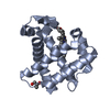
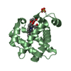
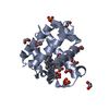
| Structure viewer | Molecule: MolmilJmol/JSmol |
|---|
- Downloads & links
-Download
| PDBx/mmCIF format | 7adq.cif.gz | 73.2 KB | Display | PDBx/mmCIF format |
|---|---|---|---|---|
| PDB format | pdb7adq.ent.gz | 52.8 KB | Display | PDB format |
| PDBx/mmJSON format | 7adq.json.gz | Tree view | PDBx/mmJSON format | |
| Others |  Other downloads Other downloads |
-Validation report
| Arichive directory | https://data.pdbj.org/pub/pdb/validation_reports/ad/7adqftp://data.pdbj.org/pub/pdb/validation_reports/ad/7adq | HTTPS FTP |
|---|
-Related structure data
| Related structure data |  7jorC  7kcuC  7kfmC  3ixfS S: Starting model for refinement C: citing same article ( |
|---|---|
| Similar structure data |


-Links
 PDBj
PDBj


- Assembly
| Deposited unit | 
| ||||||||
|---|---|---|---|---|---|---|---|---|---|
| 1 | 
| ||||||||
| 2 | 
| ||||||||
| Unit cell |
|
-Components
| #1: Protein | Mass: 15545.656 Da / Num. of mol.: 2 Source method: isolated from a genetically manipulated source Source: (gene. exp.) Amphitrite ornata (invertebrata) / Production host:  #2: Chemical |   Mass: 96.063 Da / Num. of mol.: 2 / Source method: obtained synthetically / Formula: SO4 Mass: 96.063 Da / Num. of mol.: 2 / Source method: obtained synthetically / Formula: SO4#3: Chemical |   Mass: 616.487 Da / Num. of mol.: 2 / Source method: obtained synthetically / Formula: C34H32FeN4O4 Mass: 616.487 Da / Num. of mol.: 2 / Source method: obtained synthetically / Formula: C34H32FeN4O4#4: Water | ChemComp-HOH / |  Mass: 18.015 Da / Num. of mol.: 140 / Source method: isolated from a natural source / Formula: H2O Mass: 18.015 Da / Num. of mol.: 140 / Source method: isolated from a natural source / Formula: H2OHas ligand of interest | N | Has protein modification | N | |
|---|
-Experimental details
-Experiment
| Experiment | Method: X-RAY DIFFRACTION / Number of used crystals: 1 |
|---|
- Sample preparation
| Crystal | Density Matthews: 2.22 Å3/Da / Density % sol: 44.62 % |
|---|---|
| Crystal grow | Temperature: 277 K / Method: batch mode / pH: 6 Details: 30 mg per ml DHP mixed with 200 mM ammonium sulfate 40 percent PEG 4000, in a 1 to 4 ratio in a total volume of 250 to 500 microlitres |
-Data collection
| Diffraction | Mean temperature: 293 K / Serial crystal experiment: Y | |||||||||
|---|---|---|---|---|---|---|---|---|---|---|
| Diffraction source | Source: SYNCHROTRON / Site: APS  / Beamline: 14-ID-B / Wavelength: 0.815-0.863 / Beamline: 14-ID-B / Wavelength: 0.815-0.863 | |||||||||
| Detector | Type: RAYONIX MX340-HS / Detector: CCD / Date: Apr 19, 2017 | |||||||||
| Radiation | Protocol: LAUE / Monochromatic (M) / Laue (L): L / Scattering type: x-ray / Wavelength: 0.815-0.863 | |||||||||
| Radiation wavelength |
| |||||||||
| Reflection | Resolution: 2→47.7 Å / Num. obs: 12928 / % possible obs: 67.8 % / Redundancy: 7.6 % / Rmerge(I) obs: 0.072 / Net I/σ(I): 34.1 | |||||||||
| Reflection shell | Resolution: 2→2.09 Å / Redundancy: 7.8 % / Num. unique obs: 513 / % possible all: 22 | |||||||||
| Serial crystallography sample delivery | Method: fixed target | |||||||||
| Serial crystallography sample delivery fixed target | Description: Heidelberg chip |
- Processing
| Software |
| ||||||||||||||||||||||||||||||||||||||||||
|---|---|---|---|---|---|---|---|---|---|---|---|---|---|---|---|---|---|---|---|---|---|---|---|---|---|---|---|---|---|---|---|---|---|---|---|---|---|---|---|---|---|---|---|
| Refinement | Method to determine structure: MOLECULAR REPLACEMENT Starting model: 3ixf Resolution: 2.01→47.66 Å / SU ML: 0.18 / Cross valid method: THROUGHOUT / σ(F): 0.23 / Phase error: 16.26 / Stereochemistry target values: ML
| ||||||||||||||||||||||||||||||||||||||||||
| Solvent computation | Shrinkage radii: 0.9 Å / VDW probe radii: 1.11 Å / Solvent model: FLAT BULK SOLVENT MODEL | ||||||||||||||||||||||||||||||||||||||||||
| Displacement parameters | Biso max: 49.38 Å2 / Biso mean: 15.1775 Å2 / Biso min: 1.89 Å2 | ||||||||||||||||||||||||||||||||||||||||||
| Refinement step | Cycle: final / Resolution: 2.01→47.66 Å
| ||||||||||||||||||||||||||||||||||||||||||
| LS refinement shell | Refine-ID: X-RAY DIFFRACTION / Rfactor Rfree error: 0 / Total num. of bins used: 5
|
