 Movie
Movie Controller


+ Open data

- Basic information
| Entry | Database: PDB / ID: 7acp | ||||||
|---|---|---|---|---|---|---|---|
| Title | Serial synchrotron structure of dehaloperoxidase B | ||||||
 Components Components | Dehaloperoxidase B | ||||||
 Keywords Keywords | OXIDOREDUCTASE / SSX / serial crystallography / dehaloperoxidase / peroxidase / fixed-target | ||||||
| Function / homology |  Function and homology information Function and homology informationoxygen carrier activity / peroxidase activity / oxygen binding / heme binding / metal ion binding Similarity search - Function | ||||||
| Biological species |   Amphitrite ornata (invertebrata) Amphitrite ornata (invertebrata) | ||||||
| Method |  X-RAY DIFFRACTION / SYNCHROTRON / MOLECULAR REPLACEMENT / Resolution: 1.45 Å X-RAY DIFFRACTION / SYNCHROTRON / MOLECULAR REPLACEMENT / Resolution: 1.45 Å | ||||||
 Authors Authors | Moreno Chicano, T. / Ebrahim, A. / Axford, D.A. / Sherrell, D.A. / Worrall, J.W. / Strange, R.W. / Owen, R.L. / Hough, M.A. | ||||||
| Funding support |  United Kingdom, 1items United Kingdom, 1items
| ||||||
 Citation Citation | Journal: to be published Title: SFX structure of dehaloperoxidase B in the ferric form Authors: Moreno Chicano, T. / Carey, L.M. / Ebrahim, A. / Axford, D.A. / Beale, J.H. / Sherrell, D.A. / Sugimoto, H. / Tono, K. / Owada, S. / Worrall, J.W. / Strange, R.W. / Owen, R.L. / Hough, M.A. | ||||||
| History |
|
- Structure visualization
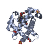
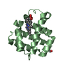
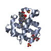
| Structure viewer | Molecule: MolmilJmol/JSmol |
|---|
- Downloads & links
-Download
| PDBx/mmCIF format | 7acp.cif.gz | 133.8 KB | Display | PDBx/mmCIF format |
|---|---|---|---|---|
| PDB format | pdb7acp.ent.gz | 104 KB | Display | PDB format |
| PDBx/mmJSON format | 7acp.json.gz | Tree view | PDBx/mmJSON format | |
| Others |  Other downloads Other downloads |
-Validation report
| Arichive directory | https://data.pdbj.org/pub/pdb/validation_reports/ac/7acpftp://data.pdbj.org/pub/pdb/validation_reports/ac/7acp | HTTPS FTP |
|---|
-Related structure data
| Related structure data |  7adfC  3ixfS S: Starting model for refinement C: citing same article ( |
|---|---|
| Similar structure data |


-Links
 PDBj
PDBj


- Assembly
| Deposited unit | 
| ||||||||
|---|---|---|---|---|---|---|---|---|---|
| 1 | 
| ||||||||
| 2 | 
| ||||||||
| Unit cell |
|
-Components
| #1: Protein | Mass: 15545.656 Da / Num. of mol.: 2 Source method: isolated from a genetically manipulated source Source: (gene. exp.) Amphitrite ornata (invertebrata) / Production host:  #2: Chemical |   Mass: 616.487 Da / Num. of mol.: 2 / Source method: obtained synthetically / Formula: C34H32FeN4O4 Mass: 616.487 Da / Num. of mol.: 2 / Source method: obtained synthetically / Formula: C34H32FeN4O4#3: Chemical |   Mass: 96.063 Da / Num. of mol.: 2 / Source method: obtained synthetically / Formula: SO4 Mass: 96.063 Da / Num. of mol.: 2 / Source method: obtained synthetically / Formula: SO4#4: Water | ChemComp-HOH / |  Mass: 18.015 Da / Num. of mol.: 173 / Source method: isolated from a natural source / Formula: H2O Mass: 18.015 Da / Num. of mol.: 173 / Source method: isolated from a natural source / Formula: H2OHas ligand of interest | N | Has protein modification | N | |
|---|
-Experimental details
-Experiment
| Experiment | Method: X-RAY DIFFRACTION / Number of used crystals: 1 |
|---|
- Sample preparation
| Crystal | Density Matthews: 2.27 Å3/Da / Density % sol: 45.86 % / Description: Microcrystals |
|---|---|
| Crystal grow | Temperature: 277 K / Method: batch mode / pH: 6 Details: Batch microcrystallization was used, mixing 30 mg/ml DHP in 20 mM MES pH 6.0 with 40%(w/v) PEG 4000, 200 mM ammonium sulfate in a 1 to 4 ratio in a total volume of 250 to 500 ml. |
-Data collection
| Diffraction | Mean temperature: 293 K / Serial crystal experiment: Y |
|---|---|
| Diffraction source | Source: SYNCHROTRON / Site: Diamond / Beamline: I24 / Wavelength: 0.9686 Å |
| Detector | Type: DECTRIS PILATUS3 6M / Detector: PIXEL / Date: Jun 28, 2018 |
| Radiation | Protocol: SINGLE WAVELENGTH / Monochromatic (M) / Laue (L): M / Scattering type: x-ray |
| Radiation wavelength | Wavelength: 0.9686 Å / Relative weight: 1 |
| Reflection | Resolution: 1.45→68.9 Å / Num. obs: 52472 / % possible obs: 100 % / Redundancy: 104 % / Biso Wilson estimate: 14.2 Å2 / CC1/2: 0.98 / R split: 0.121 / Net I/σ(I): 3.8 |
| Reflection shell | Resolution: 1.45→1.48 Å / Num. unique obs: 2504 / CC1/2: 0.977 |
| Serial crystallography sample delivery | Description: silicon 'Oxford' chip / Method: fixed target |
| Serial crystallography sample delivery fixed target | Description: silicon 'oxford' chip / Sample dehydration prevention: mylar film / Sample solvent: mother liquor |
- Processing
| Software |
| ||||||||||||||||||||||||||||||||||||||||||||||||||||||||||||||||||||||||||||||||||||||||||||||||||||||||||||||||||||||||
|---|---|---|---|---|---|---|---|---|---|---|---|---|---|---|---|---|---|---|---|---|---|---|---|---|---|---|---|---|---|---|---|---|---|---|---|---|---|---|---|---|---|---|---|---|---|---|---|---|---|---|---|---|---|---|---|---|---|---|---|---|---|---|---|---|---|---|---|---|---|---|---|---|---|---|---|---|---|---|---|---|---|---|---|---|---|---|---|---|---|---|---|---|---|---|---|---|---|---|---|---|---|---|---|---|---|---|---|---|---|---|---|---|---|---|---|---|---|---|---|---|---|
| Refinement | Method to determine structure: MOLECULAR REPLACEMENT Starting model: 3ixf Resolution: 1.45→34.466 Å / SU ML: 0.17 / Cross valid method: THROUGHOUT / σ(F): 1.35 / Phase error: 20.84 / Stereochemistry target values: ML
| ||||||||||||||||||||||||||||||||||||||||||||||||||||||||||||||||||||||||||||||||||||||||||||||||||||||||||||||||||||||||
| Solvent computation | Shrinkage radii: 0.9 Å / VDW probe radii: 1.11 Å / Solvent model: FLAT BULK SOLVENT MODEL | ||||||||||||||||||||||||||||||||||||||||||||||||||||||||||||||||||||||||||||||||||||||||||||||||||||||||||||||||||||||||
| Displacement parameters | Biso max: 134.84 Å2 / Biso mean: 26.2887 Å2 / Biso min: 9.79 Å2 | ||||||||||||||||||||||||||||||||||||||||||||||||||||||||||||||||||||||||||||||||||||||||||||||||||||||||||||||||||||||||
| Refinement step | Cycle: final / Resolution: 1.45→34.466 Å
| ||||||||||||||||||||||||||||||||||||||||||||||||||||||||||||||||||||||||||||||||||||||||||||||||||||||||||||||||||||||||
| LS refinement shell | Refine-ID: X-RAY DIFFRACTION / Rfactor Rfree error: 0
|
