 Movie
Movie Controller


+ Open data

- Basic information
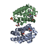
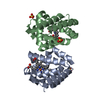
| Entry | Database: PDB / ID: 7adf | ||||||
|---|---|---|---|---|---|---|---|
| Title | SFX structure of dehaloperoxidase B in the ferric form | ||||||
 Components Components | Dehaloperoxidase B | ||||||
 Keywords Keywords | OXIDOREDUCTASE / SFX / ferric / dehaloperoxidase / peroxidase / fixed-target | ||||||
| Function / homology |  Function and homology information Function and homology informationoxygen carrier activity / peroxidase activity / oxygen binding / heme binding / metal ion binding Similarity search - Function | ||||||
| Biological species |   Amphitrite ornata (invertebrata) Amphitrite ornata (invertebrata) | ||||||
| Method |  X-RAY DIFFRACTION / FREE ELECTRON LASER / MOLECULAR REPLACEMENT / Resolution: 1.85 Å X-RAY DIFFRACTION / FREE ELECTRON LASER / MOLECULAR REPLACEMENT / Resolution: 1.85 Å | ||||||
 Authors Authors | Moreno Chicano, T. / Ebrahim, A. / Axford, D.A. / Sherrell, D.A. / Sugimoto, H. / Tono, K. / Owada, S. / Worrall, J.W. / Strange, R.W. / Owen, R.L. / Hough, M.A. | ||||||
| Funding support |  United Kingdom, 1items United Kingdom, 1items
| ||||||
 Citation Citation | Journal: to be published Title: SFX structure of dehaloperoxidase B in the ferric form Authors: Moreno Chicano, T. / Carey, L.M. / Ebrahim, A. / Axford, D.A. / Beale, J.H. / Sherrell, D.A. / Sugimoto, H. / Tono, K. / Owada, S. / Worrall, J.W. / Strange, R.W. / Owen, R.L. / Hough, M.A. | ||||||
| History |
|
- Structure visualization
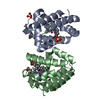
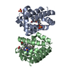
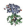
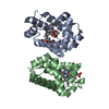
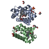
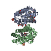
| Structure viewer | Molecule: MolmilJmol/JSmol |
|---|
- Downloads & links
-Download
| PDBx/mmCIF format | 7adf.cif.gz | 71.6 KB | Display | PDBx/mmCIF format |
|---|---|---|---|---|
| PDB format | pdb7adf.ent.gz | 51.9 KB | Display | PDB format |
| PDBx/mmJSON format | 7adf.json.gz | Tree view | PDBx/mmJSON format | |
| Others |  Other downloads Other downloads |
-Validation report
| Arichive directory | https://data.pdbj.org/pub/pdb/validation_reports/ad/7adfftp://data.pdbj.org/pub/pdb/validation_reports/ad/7adf | HTTPS FTP |
|---|
-Related structure data
| Related structure data |  7acpC  3ixfS S: Starting model for refinement C: citing same article ( |
|---|---|
| Similar structure data |

-Links
 PDBj
PDBj


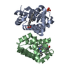
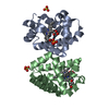
- Assembly
| Deposited unit | 
| ||||||||
|---|---|---|---|---|---|---|---|---|---|
| 1 |
| ||||||||
| Unit cell |
|
-Components
| #1: Protein | Mass: 15545.656 Da / Num. of mol.: 2 Source method: isolated from a genetically manipulated source Source: (gene. exp.) Amphitrite ornata (invertebrata) / Production host:  #2: Chemical |   Mass: 616.487 Da / Num. of mol.: 2 / Source method: obtained synthetically / Formula: C34H32FeN4O4 Mass: 616.487 Da / Num. of mol.: 2 / Source method: obtained synthetically / Formula: C34H32FeN4O4#3: Chemical |   Mass: 96.063 Da / Num. of mol.: 2 / Source method: obtained synthetically / Formula: SO4 Mass: 96.063 Da / Num. of mol.: 2 / Source method: obtained synthetically / Formula: SO4#4: Water | ChemComp-HOH / |  Mass: 18.015 Da / Num. of mol.: 84 / Source method: isolated from a natural source / Formula: H2O Mass: 18.015 Da / Num. of mol.: 84 / Source method: isolated from a natural source / Formula: H2OHas ligand of interest | N | Has protein modification | N | |
|---|
-Experimental details
-Experiment
| Experiment | Method: X-RAY DIFFRACTION / Number of used crystals: 1 |
|---|
- Sample preparation
| Crystal | Density Matthews: 2.3 Å3/Da / Density % sol: 47 % / Description: Microcrystals |
|---|---|
| Crystal grow | Temperature: 278 K / Method: batch mode / pH: 6 Details: Batch microcrystallization was used, mixing 30 mg/ml DHP in 20 mM MES pH 6.0 with 40%(w/v) PEG 4000, 200 mM ammonium sulfate in a 1 to 4 ratio in a total volume of 250 to 500 microlitres. |
-Data collection
| Diffraction | Mean temperature: 301 K / Serial crystal experiment: Y |
|---|---|
| Diffraction source | Source: FREE ELECTRON LASER / Site: SACLA  / Beamline: BL2 / Wavelength: 1.24 Å / Beamline: BL2 / Wavelength: 1.24 Å |
| Detector | Type: MPCCD / Detector: CCD / Date: Oct 12, 2017 |
| Radiation | Protocol: SINGLE WAVELENGTH / Monochromatic (M) / Laue (L): M / Scattering type: x-ray |
| Radiation wavelength | Wavelength: 1.24 Å / Relative weight: 1 |
| Reflection | Resolution: 1.85→37.9 Å / Num. obs: 25114 / % possible obs: 100 % / Redundancy: 486 % / CC1/2: 1 / R split: 0.078 / Net I/σ(I): 10.7 |
| Reflection shell | Resolution: 1.85→1.9 Å / Redundancy: 305 % / Num. unique obs: 1226 / CC1/2: 0.67 / R split: 0.603 / % possible all: 100 |
| Serial crystallography measurement | Focal spot size: 1.675 µm2 / Pulse duration: 10 fsec. / Pulse energy: 289 µJ / XFEL pulse repetition rate: 30 Hz |
| Serial crystallography sample delivery | Description: Silicon fixed target / Method: fixed target |
| Serial crystallography sample delivery fixed target | Description: Oxford Chip / Sample dehydration prevention: Mylar film |
- Processing
| Software |
| ||||||||||||||||||||||||||||||||||||||||||||||||||
|---|---|---|---|---|---|---|---|---|---|---|---|---|---|---|---|---|---|---|---|---|---|---|---|---|---|---|---|---|---|---|---|---|---|---|---|---|---|---|---|---|---|---|---|---|---|---|---|---|---|---|---|
| Refinement | Method to determine structure: MOLECULAR REPLACEMENT Starting model: PDB 3ixf Resolution: 1.85→33.955 Å / SU ML: 0.28 / Cross valid method: THROUGHOUT / σ(F): 1.36 / Phase error: 20.31 / Stereochemistry target values: ML
| ||||||||||||||||||||||||||||||||||||||||||||||||||
| Solvent computation | Shrinkage radii: 0.9 Å / VDW probe radii: 1.11 Å / Solvent model: FLAT BULK SOLVENT MODEL | ||||||||||||||||||||||||||||||||||||||||||||||||||
| Displacement parameters | Biso max: 81.51 Å2 / Biso mean: 40.0014 Å2 / Biso min: 23.81 Å2 | ||||||||||||||||||||||||||||||||||||||||||||||||||
| Refinement step | Cycle: final / Resolution: 1.85→33.955 Å
| ||||||||||||||||||||||||||||||||||||||||||||||||||
| LS refinement shell | Refine-ID: X-RAY DIFFRACTION / Rfactor Rfree error: 0 / % reflection obs: 100 %
|
