Journal: Cell / Year: 2022 Title: BacPROTACs mediate targeted protein degradation in bacteria. Authors: Francesca E Morreale / Stefan Kleine / Julia Leodolter / Sabryna Junker / David M Hoi / Stepan Ovchinnikov / Anastasia Okun / Juliane Kley / Robert Kurzbauer / Lukas Junk / Somraj Guha / ...Authors: Francesca E Morreale / Stefan Kleine / Julia Leodolter / Sabryna Junker / David M Hoi / Stepan Ovchinnikov / Anastasia Okun / Juliane Kley / Robert Kurzbauer / Lukas Junk / Somraj Guha / David Podlesainski / Uli Kazmaier / Guido Boehmelt / Harald Weinstabl / Klaus Rumpel / Volker M Schmiedel / Markus Hartl / David Haselbach / Anton Meinhart / Markus Kaiser / Tim Clausen / Abstract: Hijacking the cellular protein degradation system offers unique opportunities for drug discovery, as exemplified by proteolysis-targeting chimeras. Despite their great promise for medical chemistry, ...Hijacking the cellular protein degradation system offers unique opportunities for drug discovery, as exemplified by proteolysis-targeting chimeras. Despite their great promise for medical chemistry, so far, it has not been possible to reprogram the bacterial degradation machinery to interfere with microbial infections. Here, we develop small-molecule degraders, so-called BacPROTACs, that bind to the substrate receptor of the ClpC:ClpP protease, priming neo-substrates for degradation. In addition to their targeting function, BacPROTACs activate ClpC, transforming the resting unfoldase into its functional state. The induced higher-order oligomer was visualized by cryo-EM analysis, providing a structural snapshot of activated ClpC unfolding a protein substrate. Finally, drug susceptibility and degradation assays performed in mycobacteria demonstrate in vivo activity of BacPROTACs, allowing selective targeting of endogenous proteins via fusion to an established degron. In addition to guiding antibiotic discovery, the BacPROTAC technology presents a versatile research tool enabling the inducible degradation of bacterial proteins.
In the structure databanks used in Yorodumi, some data are registered as the other names, "COVID-19 virus" and "2019-nCoV". Here are the details of the virus and the list of structure data.
Jan 31, 2019. EMDB accession codes are about to change! (news from PDBe EMDB page)
EMDB accession codes are about to change! (news from PDBe EMDB page)
The allocation of 4 digits for EMDB accession codes will soon come to an end. Whilst these codes will remain in use, new EMDB accession codes will include an additional digit and will expand incrementally as the available range of codes is exhausted. The current 4-digit format prefixed with “EMD-” (i.e. EMD-XXXX) will advance to a 5-digit format (i.e. EMD-XXXXX), and so on. It is currently estimated that the 4-digit codes will be depleted around Spring 2019, at which point the 5-digit format will come into force.
The EM Navigator/Yorodumi systems omit the EMD- prefix.
Related info.:Q: What is EMD? / ID/Accession-code notation in Yorodumi/EM Navigator
Yorodumi is a browser for structure data from EMDB, PDB, SASBDB, etc.
This page is also the successor to EM Navigator detail page, and also detail information page/front-end page for Omokage search.
The word "yorodu" (or yorozu) is an old Japanese word meaning "ten thousand". "mi" (miru) is to see.
Related info.:EMDB / PDB / SASBDB / Comparison of 3 databanks / Yorodumi Search / Aug 31, 2016. New EM Navigator & Yorodumi / Yorodumi Papers / Jmol/JSmol / Function and homology information / Changes in new EM Navigator and Yorodumi
 Movie
Movie Controller


 Open data
 Basic information
 Components
 Keywords
 Function and homology information
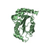
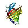
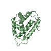
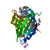
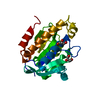
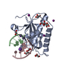
 Mycobacterium tuberculosis (bacteria)
 X-RAY DIFFRACTION /
  Authors
 Austria, 1items
  Citation
 Structure visualization
 Downloads & links
 Other downloads
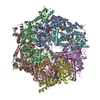


 PDBj
PDBj
 Assembly
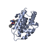
 Mass: 18.015 Da / Num. of mol.: 192 / Source method: isolated from a natural source / Formula: H2O
 Sample preparation
 Processing