 Movie
Movie Controller

[English] 日本語
 Yorodumi
- PDB-7a99: Crystal structure of the Phe57Trp mutant of the arginine-bound fo... -
+ Open data

- Basic information
| Entry | Database: PDB / ID: 7a99 | ||||||
|---|---|---|---|---|---|---|---|
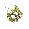
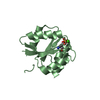
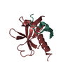
| Title | Crystal structure of the Phe57Trp mutant of the arginine-bound form of domain 1 from TmArgBP | ||||||
 Components Components | Amino acid ABC transporter, periplasmic amino acid-binding protein,Amino acid ABC transporter, periplasmic amino acid-binding protein | ||||||
 Keywords Keywords | TRANSPORT PROTEIN / Arginine ligand / Arginine sensing / protein dissection | ||||||
| Function / homology |  Function and homology information Function and homology informationamino acid binding / ligand-gated monoatomic ion channel activity / outer membrane-bounded periplasmic space / membrane Similarity search - Function | ||||||
| Biological species |   Thermotoga maritima (bacteria) Thermotoga maritima (bacteria) | ||||||
| Method |  X-RAY DIFFRACTION / MOLECULAR REPLACEMENT / Resolution: 1.79 Å X-RAY DIFFRACTION / MOLECULAR REPLACEMENT / Resolution: 1.79 Å | ||||||
 Authors Authors | Balasco, N. / Vitagliano, L. / Smaldone, G. / Ruggiero, A. | ||||||
 Citation Citation | Journal: Int J Mol Sci / Year: 2020 Title: Development of a Protein Scaffold for Arginine Sensing Generated through the Dissection of the Arginine-Binding Protein from Thermotoga maritima . Authors: Smaldone, G. / Ruggiero, A. / Balasco, N. / Vitagliano, L. | ||||||
| History |
|
- Structure visualization
| Structure viewer | Molecule: MolmilJmol/JSmol |
|---|
- Downloads & links
-Download
| PDBx/mmCIF format | 7a99.cif.gz | 71.9 KB | Display | PDBx/mmCIF format |
|---|---|---|---|---|
| PDB format | pdb7a99.ent.gz | 52.4 KB | Display | PDB format |
| PDBx/mmJSON format | 7a99.json.gz | Tree view | PDBx/mmJSON format | |
| Others |  Other downloads Other downloads |
-Validation report
| Arichive directory | https://data.pdbj.org/pub/pdb/validation_reports/a9/7a99ftp://data.pdbj.org/pub/pdb/validation_reports/a9/7a99 | HTTPS FTP |
|---|
-Related structure data
| Related structure data |  6gpcS S: Starting model for refinement |
|---|---|
| Similar structure data |


-Links
 PDBj
PDBj

- Assembly
| Deposited unit | 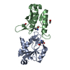
| ||||||||
|---|---|---|---|---|---|---|---|---|---|
| 1 | 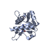
| ||||||||
| 2 | 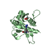
| ||||||||
| Unit cell |
|
-Components
| #1: Protein | Mass: 15226.337 Da / Num. of mol.: 2 Source method: isolated from a genetically manipulated source Source: (gene. exp.) Thermotoga maritima (bacteria), (gene. exp.) Thermotoga maritima (strain ATCC 43589 / MSB8 / DSM 3109 / JCM 10099) (bacteria)Gene: TM_0593 / Strain: ATCC 43589 / MSB8 / DSM 3109 / JCM 10099 / Production host: #2: Chemical |   Type: L-peptide linking / Mass: 175.209 Da / Num. of mol.: 2 / Source method: obtained synthetically / Formula: C6H15N4O2 / Feature type: SUBJECT OF INVESTIGATION Type: L-peptide linking / Mass: 175.209 Da / Num. of mol.: 2 / Source method: obtained synthetically / Formula: C6H15N4O2 / Feature type: SUBJECT OF INVESTIGATION#3: Chemical |   Mass: 59.044 Da / Num. of mol.: 3 / Source method: obtained synthetically / Formula: C2H3O2 Mass: 59.044 Da / Num. of mol.: 3 / Source method: obtained synthetically / Formula: C2H3O2#4: Water | ChemComp-HOH / |  Mass: 18.015 Da / Num. of mol.: 297 / Source method: isolated from a natural source / Formula: H2O Mass: 18.015 Da / Num. of mol.: 297 / Source method: isolated from a natural source / Formula: H2OHas ligand of interest | Y | |
|---|
-Experimental details
-Experiment
| Experiment | Method: X-RAY DIFFRACTION / Number of used crystals: 1 |
|---|
- Sample preparation
| Crystal | Density Matthews: 2.12 Å3/Da / Density % sol: 41.93 % |
|---|---|
| Crystal grow | Temperature: 293 K / Method: vapor diffusion, hanging drop Details: 0.1 M Sodium acetate trihydrate pH 4.5, 25% w/v Polyethylene glycol 3,350 |
-Data collection
| Diffraction | Mean temperature: 100 K / Serial crystal experiment: N |
|---|---|
| Diffraction source | Source: ROTATING ANODE / Type: RIGAKU MICROMAX-007 HF / Wavelength: 1.54 Å |
| Detector | Type: RIGAKU SATURN 944 / Detector: CCD / Date: May 11, 2016 |
| Radiation | Protocol: SINGLE WAVELENGTH / Monochromatic (M) / Laue (L): M / Scattering type: x-ray |
| Radiation wavelength | Wavelength: 1.54 Å / Relative weight: 1 |
| Reflection | Resolution: 1.79→56.19 Å / Num. obs: 25021 / % possible obs: 99.1 % / Redundancy: 4 % / Rpim(I) all: 0.033 / Net I/σ(I): 27.4 |
| Reflection shell | Resolution: 1.79→1.87 Å / Num. unique obs: 3013 / Rpim(I) all: 0.317 |
- Processing
| Software |
| ||||||||||||||||||||||||||||||||||||||||||||||||||||||||||||
|---|---|---|---|---|---|---|---|---|---|---|---|---|---|---|---|---|---|---|---|---|---|---|---|---|---|---|---|---|---|---|---|---|---|---|---|---|---|---|---|---|---|---|---|---|---|---|---|---|---|---|---|---|---|---|---|---|---|---|---|---|---|
| Refinement | Method to determine structure: MOLECULAR REPLACEMENT Starting model: 6GPC Resolution: 1.79→56.19 Å / Cor.coef. Fo:Fc: 0.963 / Cor.coef. Fo:Fc free: 0.936 / SU B: 2.95 / SU ML: 0.091 / Cross valid method: THROUGHOUT / σ(F): 0 / ESU R: 0.132 / ESU R Free: 0.134 / Stereochemistry target values: MAXIMUM LIKELIHOOD Details: HYDROGENS HAVE BEEN ADDED IN THE RIDING POSITIONS U VALUES : REFINED INDIVIDUALLY
| ||||||||||||||||||||||||||||||||||||||||||||||||||||||||||||
| Solvent computation | Ion probe radii: 0.8 Å / Shrinkage radii: 0.8 Å / VDW probe radii: 1.2 Å / Solvent model: MASK | ||||||||||||||||||||||||||||||||||||||||||||||||||||||||||||
| Displacement parameters | Biso max: 79.53 Å2 / Biso mean: 24.745 Å2 / Biso min: 12.1 Å2
| ||||||||||||||||||||||||||||||||||||||||||||||||||||||||||||
| Refinement step | Cycle: final / Resolution: 1.79→56.19 Å
| ||||||||||||||||||||||||||||||||||||||||||||||||||||||||||||
| Refine LS restraints |
| ||||||||||||||||||||||||||||||||||||||||||||||||||||||||||||
| LS refinement shell | Resolution: 1.79→1.834 Å / Rfactor Rfree error: 0
|
