 Movie
Movie Controller

[English] 日本語
 Yorodumi
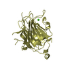
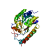
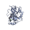
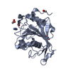
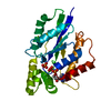
- PDB-7a8v: Crystal structure of Polysaccharide monooxygenase from P.verruculosum -
+ Open data

- Basic information
| Entry | Database: PDB / ID: 7a8v | ||||||
|---|---|---|---|---|---|---|---|
| Title | Crystal structure of Polysaccharide monooxygenase from P.verruculosum | ||||||
 Components Components | Lytic polysaccharide monooxygenase | ||||||
 Keywords Keywords | OXIDOREDUCTASE / MONOOXYGENASE / P.verruculosum | ||||||
| Function / homology |  Function and homology information Function and homology informationlytic cellulose monooxygenase (C4-dehydrogenating) / hydrolase activity, acting on glycosyl bonds / cellulose catabolic process / monooxygenase activity / extracellular region Similarity search - Function | ||||||
| Biological species |  Talaromyces verruculosus (fungus) Talaromyces verruculosus (fungus) | ||||||
| Method |  X-RAY DIFFRACTION / MOLECULAR REPLACEMENT / Resolution: 1.94961087137 Å X-RAY DIFFRACTION / MOLECULAR REPLACEMENT / Resolution: 1.94961087137 Å | ||||||
 Authors Authors | Nemashkalov, V. / Kravchenko, O. / Gabdulkhakov, A. / Tischenko, S. / Rozhkova, A. / Sinitsyn, A. | ||||||
| Funding support | 1items
| ||||||
 Citation Citation | Journal: To Be Published Title: Crystal structure of Polysaccharide monooxygenase from P.verruculosum Authors: Nemashkalov, V. / Kravchenko, O. / Gabdulkhakov, A. / Tischenko, S. / Rozhkova, A. / Sinitsyn, A. | ||||||
| History |
|
- Structure visualization
| Structure viewer | Molecule: MolmilJmol/JSmol |
|---|
- Downloads & links
-Download
| PDBx/mmCIF format | 7a8v.cif.gz | 69 KB | Display | PDBx/mmCIF format |
|---|---|---|---|---|
| PDB format | pdb7a8v.ent.gz | 47.6 KB | Display | PDB format |
| PDBx/mmJSON format | 7a8v.json.gz | Tree view | PDBx/mmJSON format | |
| Others |  Other downloads Other downloads |
-Validation report
| Arichive directory | https://data.pdbj.org/pub/pdb/validation_reports/a8/7a8vftp://data.pdbj.org/pub/pdb/validation_reports/a8/7a8v | HTTPS FTP |
|---|
-Related structure data
| Related structure data |  4eisS S: Starting model for refinement |
|---|---|
| Similar structure data |


-Links
 PDBj
PDBj


- Assembly
| Deposited unit | 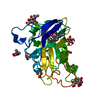
| ||||||||||||
|---|---|---|---|---|---|---|---|---|---|---|---|---|---|
| 1 |
| ||||||||||||
| Unit cell |
| ||||||||||||
| Components on special symmetry positions |
|
-Components
-Protein , 1 types, 1 molecules A
| #1: Protein | Mass: 24367.729 Da / Num. of mol.: 1 Source method: isolated from a genetically manipulated source Source: (gene. exp.) Talaromyces verruculosus (fungus) / Gene: lpmo1 / Production host: Penicillium canescens (fungus) / References: UniProt: A0A482A9N4 |
|---|
-Sugars , 2 types, 8 molecules 

| #2: Sugar |  Type: D-saccharide, beta linking / Mass: 221.208 Da / Num. of mol.: 3 / Source method: obtained synthetically / Formula: C8H15NO6 / Feature type: SUBJECT OF INVESTIGATION Type: D-saccharide, beta linking / Mass: 221.208 Da / Num. of mol.: 3 / Source method: obtained synthetically / Formula: C8H15NO6 / Feature type: SUBJECT OF INVESTIGATION#3: Sugar | ChemComp-MAN /  Type: D-saccharide, alpha linking / Mass: 180.156 Da / Num. of mol.: 5 / Source method: obtained synthetically / Formula: C6H12O6 / Feature type: SUBJECT OF INVESTIGATION Type: D-saccharide, alpha linking / Mass: 180.156 Da / Num. of mol.: 5 / Source method: obtained synthetically / Formula: C6H12O6 / Feature type: SUBJECT OF INVESTIGATION |
|---|
-Non-polymers , 3 types, 261 molecules 


| #4: Chemical |  Mass: 96.063 Da / Num. of mol.: 3 / Source method: obtained synthetically / Formula: SO4 / Feature type: SUBJECT OF INVESTIGATION Mass: 96.063 Da / Num. of mol.: 3 / Source method: obtained synthetically / Formula: SO4 / Feature type: SUBJECT OF INVESTIGATION#5: Chemical | ChemComp-CU / |  Mass: 63.546 Da / Num. of mol.: 1 / Source method: obtained synthetically / Formula: Cu / Feature type: SUBJECT OF INVESTIGATION Mass: 63.546 Da / Num. of mol.: 1 / Source method: obtained synthetically / Formula: Cu / Feature type: SUBJECT OF INVESTIGATION#6: Water | ChemComp-HOH / | Mass: 18.015 Da / Num. of mol.: 257 / Source method: isolated from a natural source / Formula: H2O |
|---|
-Details
| Has ligand of interest | Y |
|---|---|
| Has protein modification | Y |
-Experimental details
-Experiment
| Experiment | Method: X-RAY DIFFRACTION / Number of used crystals: 1 |
|---|
- Sample preparation
| Crystal | Density Matthews: 4.13 Å3/Da / Density % sol: 70.24 % |
|---|---|
| Crystal grow | Temperature: 295 K / Method: vapor diffusion, hanging drop / Details: cobalt chloride, MES, ammonium sulfate |
-Data collection
| Diffraction | Mean temperature: 110 K / Serial crystal experiment: N |
|---|---|
| Diffraction source | Source: ROTATING ANODE / Type: BRUKER AXS MICROSTAR / Wavelength: 1.54 Å |
| Detector | Type: Bruker Platinum 135 / Detector: CCD / Date: Apr 16, 2020 |
| Radiation | Protocol: SINGLE WAVELENGTH / Monochromatic (M) / Laue (L): M / Scattering type: x-ray |
| Radiation wavelength | Wavelength: 1.54 Å / Relative weight: 1 |
| Reflection | Resolution: 1.94→33.04 Å / Num. obs: 29254 / % possible obs: 99 % / Redundancy: 4.04 % / Biso Wilson estimate: 10.7624280739 Å2 / Rmerge(I) obs: 0.1038 / Net I/σ(I): 10.28 |
| Reflection shell | Resolution: 1.95→2.05 Å / Rmerge(I) obs: 0.2844 / Num. unique obs: 3860 |
- Processing
| Software |
| |||||||||||||||||||||||||||||||||||||||||||||||||||||||||||||||||||||||||||||
|---|---|---|---|---|---|---|---|---|---|---|---|---|---|---|---|---|---|---|---|---|---|---|---|---|---|---|---|---|---|---|---|---|---|---|---|---|---|---|---|---|---|---|---|---|---|---|---|---|---|---|---|---|---|---|---|---|---|---|---|---|---|---|---|---|---|---|---|---|---|---|---|---|---|---|---|---|---|---|
| Refinement | Method to determine structure: MOLECULAR REPLACEMENT Starting model: 4EIS Resolution: 1.94961087137→33.03625 Å / SU ML: 0.173506366293 / Cross valid method: FREE R-VALUE / σ(F): 1.34760157227 / Phase error: 16.3330556472 Stereochemistry target values: GeoStd + Monomer Library + CDL v1.2
| |||||||||||||||||||||||||||||||||||||||||||||||||||||||||||||||||||||||||||||
| Solvent computation | Shrinkage radii: 0.9 Å / VDW probe radii: 1.11 Å / Solvent model: FLAT BULK SOLVENT MODEL | |||||||||||||||||||||||||||||||||||||||||||||||||||||||||||||||||||||||||||||
| Displacement parameters | Biso mean: 13.3679876308 Å2 | |||||||||||||||||||||||||||||||||||||||||||||||||||||||||||||||||||||||||||||
| Refinement step | Cycle: LAST / Resolution: 1.94961087137→33.03625 Å
| |||||||||||||||||||||||||||||||||||||||||||||||||||||||||||||||||||||||||||||
| Refine LS restraints |
| |||||||||||||||||||||||||||||||||||||||||||||||||||||||||||||||||||||||||||||
| LS refinement shell |
|
