 Movie
Movie Controller

[English] 日本語
 Yorodumi
- PDB-7a6y: Structure of 14-3-3 gamma in complex with DAPK2 peptide stabilize... -
+ Open data

- Basic information
| Entry | Database: PDB / ID: 7a6y | ||||||
|---|---|---|---|---|---|---|---|
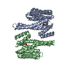
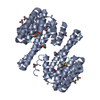
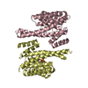
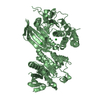
| Title | Structure of 14-3-3 gamma in complex with DAPK2 peptide stabilized by FC-A | ||||||
 Components Components |
| ||||||
 Keywords Keywords | SIGNALING PROTEIN / 14-3-3 protein / DAPK2 / kinase / complex / phosphorylation / fusicoccin | ||||||
| Function / homology |  Function and homology information Function and homology informationpositive regulation of cell-cell adhesion / phosphorylation-dependent protein binding / positive regulation of T cell mediated immune response to tumor cell / regulation of neuron differentiation / protein kinase C inhibitor activity / Regulation of localization of FOXO transcription factors / Activation of BAD and translocation to mitochondria / regulation of signal transduction / SARS-CoV-2 targets host intracellular signalling and regulatory pathways / negative regulation of protein kinase activity ...positive regulation of cell-cell adhesion / phosphorylation-dependent protein binding / positive regulation of T cell mediated immune response to tumor cell / regulation of neuron differentiation / protein kinase C inhibitor activity / Regulation of localization of FOXO transcription factors / Activation of BAD and translocation to mitochondria / regulation of signal transduction / SARS-CoV-2 targets host intracellular signalling and regulatory pathways / negative regulation of protein kinase activity / protein targeting / cellular response to glucose starvation / SARS-CoV-1 targets host intracellular signalling and regulatory pathways / RHO GTPases activate PKNs / Chk1/Chk2(Cds1) mediated inactivation of Cyclin B:Cdk1 complex / insulin-like growth factor receptor binding / negative regulation of TORC1 signaling / Loss of Nlp from mitotic centrosomes / Loss of proteins required for interphase microtubule organization from the centrosome / Transcriptional and post-translational regulation of MITF-M expression and activity / Recruitment of mitotic centrosome proteins and complexes / Recruitment of NuMA to mitotic centrosomes / Anchoring of the basal body to the plasma membrane / AURKA Activation by TPX2 / protein kinase C binding / TP53 Regulates Metabolic Genes / Translocation of SLC2A4 (GLUT4) to the plasma membrane / protein sequestering activity / receptor tyrosine kinase binding / regulation of synaptic plasticity / positive regulation of T cell activation / cellular response to insulin stimulus / intracellular protein localization / Regulation of PLK1 Activity at G2/M Transition / regulation of protein localization / presynapse / mitochondrial matrix / protein domain specific binding / focal adhesion / signal transduction / RNA binding / extracellular exosome / membrane / identical protein binding / nucleus / cytoplasm / cytosol Similarity search - Function | ||||||
| Biological species |  Homo sapiens (human) Homo sapiens (human) | ||||||
| Method |  X-RAY DIFFRACTION / MOLECULAR REPLACEMENT / Resolution: 2.5 Å X-RAY DIFFRACTION / MOLECULAR REPLACEMENT / Resolution: 2.5 Å | ||||||
 Authors Authors | Horvath, M. / Obsilova, V. / Obsil, T. | ||||||
| Funding support |  Czech Republic, 1items Czech Republic, 1items
| ||||||
 Citation Citation | Journal: Commun Biol / Year: 2021 Title: 14-3-3 proteins inactivate DAPK2 by promoting its dimerization and protecting key regulatory phosphosites. Authors: Horvath, M. / Petrvalska, O. / Herman, P. / Obsilova, V. / Obsil, T. | ||||||
| History |
|
- Structure visualization
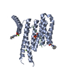
| Structure viewer | Molecule: MolmilJmol/JSmol |
|---|
- Downloads & links
-Download
| PDBx/mmCIF format | 7a6y.cif.gz | 241.4 KB | Display | PDBx/mmCIF format |
|---|---|---|---|---|
| PDB format | pdb7a6y.ent.gz | 157.2 KB | Display | PDB format |
| PDBx/mmJSON format | 7a6y.json.gz | Tree view | PDBx/mmJSON format | |
| Others |  Other downloads Other downloads |
-Validation report
| Arichive directory | https://data.pdbj.org/pub/pdb/validation_reports/a6/7a6yftp://data.pdbj.org/pub/pdb/validation_reports/a6/7a6y | HTTPS FTP |
|---|
-Related structure data
| Related structure data |  7a6rC 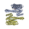 2b05S S: Starting model for refinement C: citing same article ( |
|---|---|
| Similar structure data |


-Links
 PDBj
PDBj


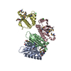
- Assembly
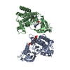
| Deposited unit | 
| ||||||||||||
|---|---|---|---|---|---|---|---|---|---|---|---|---|---|
| 1 | 
| ||||||||||||
| 2 | 
| ||||||||||||
| Unit cell |
|
-Components
| #1: Protein | Mass: 27199.625 Da / Num. of mol.: 4 Source method: isolated from a genetically manipulated source Source: (gene. exp.) Homo sapiens (human) / Gene: YWHAG / Production host:  #2: Protein/peptide | Mass: 931.914 Da / Num. of mol.: 4 / Source method: obtained synthetically / Source: (synth.) Homo sapiens (human)#3: Chemical | 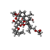  Mass: 680.823 Da / Num. of mol.: 3 / Source method: obtained synthetically / Formula: C36H56O12 Mass: 680.823 Da / Num. of mol.: 3 / Source method: obtained synthetically / Formula: C36H56O12#4: Water | ChemComp-HOH / |  Mass: 18.015 Da / Num. of mol.: 151 / Source method: isolated from a natural source / Formula: H2O Mass: 18.015 Da / Num. of mol.: 151 / Source method: isolated from a natural source / Formula: H2OHas ligand of interest | N | Has protein modification | Y | |
|---|
-Experimental details
-Experiment
| Experiment | Method: X-RAY DIFFRACTION / Number of used crystals: 1 |
|---|
- Sample preparation
| Crystal | Density Matthews: 2.68 Å3/Da / Density % sol: 54.14 % |
|---|---|
| Crystal grow | Temperature: 290.15 K / Method: vapor diffusion, hanging drop / pH: 7.5 / Details: HEPES, MgCl2, PEG400, hexafluoro-2-propanol, FC-A |
-Data collection
| Diffraction | Mean temperature: 100 K / Serial crystal experiment: N |
|---|---|
| Diffraction source | Source: ROTATING ANODE / Type: RIGAKU MICROMAX-007 HF / Wavelength: 1.54187 Å |
| Detector | Type: DECTRIS PILATUS 300K / Detector: PIXEL / Date: Mar 13, 2019 |
| Radiation | Protocol: SINGLE WAVELENGTH / Monochromatic (M) / Laue (L): M / Scattering type: x-ray |
| Radiation wavelength | Wavelength: 1.54187 Å / Relative weight: 1 |
| Reflection | Resolution: 2.5→29.68 Å / Num. obs: 40349 / % possible obs: 99.64 % / Redundancy: 4.23 % / Biso Wilson estimate: 45.12 Å2 / CC1/2: 0.999 / Rmerge(I) obs: 0.036 / Rrim(I) all: 0.041 / Net I/σ(I): 26.4 |
| Reflection shell | Resolution: 2.5→2.589 Å / Rmerge(I) obs: 0.219 / Mean I/σ(I) obs: 3.6 / Num. unique obs: 4042 / CC1/2: 0.916 / Rrim(I) all: 0.292 / % possible all: 99.65 |
- Processing
| Software |
| |||||||||||||||||||||||||||||||||||||||||||||||||||||||||||||||||||||||||||||||||||||||||||||||||||||||||||||||||||||||||||||||||||||||||||||||||||||||||||||||||||||||||||||||||||||||||||||
|---|---|---|---|---|---|---|---|---|---|---|---|---|---|---|---|---|---|---|---|---|---|---|---|---|---|---|---|---|---|---|---|---|---|---|---|---|---|---|---|---|---|---|---|---|---|---|---|---|---|---|---|---|---|---|---|---|---|---|---|---|---|---|---|---|---|---|---|---|---|---|---|---|---|---|---|---|---|---|---|---|---|---|---|---|---|---|---|---|---|---|---|---|---|---|---|---|---|---|---|---|---|---|---|---|---|---|---|---|---|---|---|---|---|---|---|---|---|---|---|---|---|---|---|---|---|---|---|---|---|---|---|---|---|---|---|---|---|---|---|---|---|---|---|---|---|---|---|---|---|---|---|---|---|---|---|---|---|---|---|---|---|---|---|---|---|---|---|---|---|---|---|---|---|---|---|---|---|---|---|---|---|---|---|---|---|---|---|---|---|---|
| Refinement | Method to determine structure: MOLECULAR REPLACEMENT Starting model: 2B05 Resolution: 2.5→29.68 Å / SU ML: 0.3185 / Cross valid method: FREE R-VALUE / σ(F): 1.16 / Phase error: 30.9377 Stereochemistry target values: GeoStd + Monomer Library + CDL v1.2
| |||||||||||||||||||||||||||||||||||||||||||||||||||||||||||||||||||||||||||||||||||||||||||||||||||||||||||||||||||||||||||||||||||||||||||||||||||||||||||||||||||||||||||||||||||||||||||||
| Solvent computation | Shrinkage radii: 0.9 Å / VDW probe radii: 1.11 Å / Solvent model: FLAT BULK SOLVENT MODEL | |||||||||||||||||||||||||||||||||||||||||||||||||||||||||||||||||||||||||||||||||||||||||||||||||||||||||||||||||||||||||||||||||||||||||||||||||||||||||||||||||||||||||||||||||||||||||||||
| Displacement parameters | Biso mean: 52.32 Å2 | |||||||||||||||||||||||||||||||||||||||||||||||||||||||||||||||||||||||||||||||||||||||||||||||||||||||||||||||||||||||||||||||||||||||||||||||||||||||||||||||||||||||||||||||||||||||||||||
| Refinement step | Cycle: LAST / Resolution: 2.5→29.68 Å
| |||||||||||||||||||||||||||||||||||||||||||||||||||||||||||||||||||||||||||||||||||||||||||||||||||||||||||||||||||||||||||||||||||||||||||||||||||||||||||||||||||||||||||||||||||||||||||||
| Refine LS restraints |
| |||||||||||||||||||||||||||||||||||||||||||||||||||||||||||||||||||||||||||||||||||||||||||||||||||||||||||||||||||||||||||||||||||||||||||||||||||||||||||||||||||||||||||||||||||||||||||||
| LS refinement shell |
|
