 Movie
Movie Controller


+ Open data

- Basic information
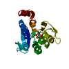
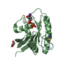
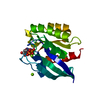
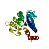
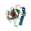
| Entry | Database: PDB / ID: 7a47 | ||||||
|---|---|---|---|---|---|---|---|
| Title | KRASG12C GDP form in complex with Cpd4 | ||||||
 Components Components | Isoform 2B of GTPase KRas | ||||||
 Keywords Keywords | ONCOPROTEIN / inhibitor / mutant | ||||||
| Function / homology | small monomeric GTPase / Ca2+ pathway / P-loop containing nucleotide triphosphate hydrolases / Rossmann fold / 3-Layer(aba) Sandwich / Alpha Beta / GUANOSINE-5'-DIPHOSPHATE / Chem-QY5 / Isoform 2B of GTPase KRas Function and homology information Function and homology information | ||||||
| Biological species |  Homo sapiens (human) Homo sapiens (human) | ||||||
| Method |  X-RAY DIFFRACTION / SYNCHROTRON / MOLECULAR REPLACEMENT / Resolution: 2.16 Å X-RAY DIFFRACTION / SYNCHROTRON / MOLECULAR REPLACEMENT / Resolution: 2.16 Å | ||||||
 Authors Authors | Bertrand, T. / Steier, V. | ||||||
 Citation Citation | Journal: Small Gtpases / Year: 2022 Title: KRAS G12C fragment screening renders new binding pockets. Authors: Mathieu, M. / Steier, V. / Fassy, F. / Delorme, C. / Papin, D. / Genet, B. / Duffieux, F. / Bertrand, T. / Delarbre, L. / Le-Borgne, H. / Parent, A. / Didier, P. / Marquette, J.P. / ...Authors: Mathieu, M. / Steier, V. / Fassy, F. / Delorme, C. / Papin, D. / Genet, B. / Duffieux, F. / Bertrand, T. / Delarbre, L. / Le-Borgne, H. / Parent, A. / Didier, P. / Marquette, J.P. / Lowinski, M. / Houtmann, J. / Lamberton, A. / Debussche, L. / Alexey, R. | ||||||
| History |
|
- Structure visualization
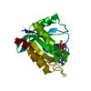
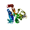
| Structure viewer | Molecule: MolmilJmol/JSmol |
|---|
- Downloads & links
-Download
| PDBx/mmCIF format | 7a47.cif.gz | 121.2 KB | Display | PDBx/mmCIF format |
|---|---|---|---|---|
| PDB format | pdb7a47.ent.gz | 94.9 KB | Display | PDB format |
| PDBx/mmJSON format | 7a47.json.gz | Tree view | PDBx/mmJSON format | |
| Others |  Other downloads Other downloads |
-Validation report
| Arichive directory | https://data.pdbj.org/pub/pdb/validation_reports/a4/7a47ftp://data.pdbj.org/pub/pdb/validation_reports/a4/7a47 | HTTPS FTP |
|---|
-Related structure data
| Related structure data |  7a1wC  7a1xSC  7a1yC S: Starting model for refinement C: citing same article ( |
|---|---|
| Similar structure data |


-Links
 PDBj
PDBj


- Assembly
| Deposited unit | 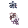
| ||||||||
|---|---|---|---|---|---|---|---|---|---|
| 1 | 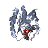
| ||||||||
| 2 | 
| ||||||||
| 3 | 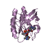
| ||||||||
| Unit cell |
|
-Components
| #1: Protein | Mass: 19374.902 Da / Num. of mol.: 3 / Mutation: G12C Source method: isolated from a genetically manipulated source Details: G12C mutant / Source: (gene. exp.) Homo sapiens (human) / Gene: KRAS, KRAS2, RASK2 / Plasmid: pET-28a / Details (production host): T7 promoter / Production host:  #2: Chemical |   Type: RNA linking / Mass: 443.201 Da / Num. of mol.: 3 / Source method: obtained synthetically / Formula: C10H15N5O11P2 / Comment: GDP, energy-carrying molecule*YM Type: RNA linking / Mass: 443.201 Da / Num. of mol.: 3 / Source method: obtained synthetically / Formula: C10H15N5O11P2 / Comment: GDP, energy-carrying molecule*YM#3: Chemical | 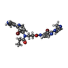  Mass: 616.308 Da / Num. of mol.: 3 / Source method: obtained synthetically / Formula: C24H24Br2N8O2 / Feature type: SUBJECT OF INVESTIGATION Mass: 616.308 Da / Num. of mol.: 3 / Source method: obtained synthetically / Formula: C24H24Br2N8O2 / Feature type: SUBJECT OF INVESTIGATION#4: Water | ChemComp-HOH / |  Mass: 18.015 Da / Num. of mol.: 211 / Source method: isolated from a natural source / Formula: H2O Mass: 18.015 Da / Num. of mol.: 211 / Source method: isolated from a natural source / Formula: H2OHas ligand of interest | Y | Has protein modification | Y | |
|---|
-Experimental details
-Experiment
| Experiment | Method: X-RAY DIFFRACTION / Number of used crystals: 1 |
|---|
- Sample preparation
| Crystal | Density Matthews: 3.54 Å3/Da / Density % sol: 65.21 % |
|---|---|
| Crystal grow | Temperature: 293 K / Method: vapor diffusion, hanging drop / pH: 6 / Details: 1.1 M Sodium Citrate pH 6.0 |
-Data collection
| Diffraction | Mean temperature: 100 K / Serial crystal experiment: N |
|---|---|
| Diffraction source | Source: SYNCHROTRON / Site: ESRF  / Beamline: ID23-1 / Wavelength: 0.976256 Å / Beamline: ID23-1 / Wavelength: 0.976256 Å |
| Detector | Type: DECTRIS PILATUS3 6M / Detector: PIXEL / Date: Oct 8, 2014 |
| Radiation | Protocol: SINGLE WAVELENGTH / Monochromatic (M) / Laue (L): M / Scattering type: x-ray |
| Radiation wavelength | Wavelength: 0.976256 Å / Relative weight: 1 |
| Reflection | Resolution: 2.16→30.73 Å / Num. obs: 44756 / % possible obs: 99.7 % / Redundancy: 9.9 % / Biso Wilson estimate: 40.59 Å2 / Rmerge(I) obs: 0.122 / Net I/σ(I): 13.9 |
| Reflection shell | Resolution: 2.16→2.22 Å / Rmerge(I) obs: 0.688 / Mean I/σ(I) obs: 3 / Num. unique obs: 3443 |
- Processing
| Software |
| ||||||||||||||||||||||||||||||||||||||||||||||||||||||||||||||||||
|---|---|---|---|---|---|---|---|---|---|---|---|---|---|---|---|---|---|---|---|---|---|---|---|---|---|---|---|---|---|---|---|---|---|---|---|---|---|---|---|---|---|---|---|---|---|---|---|---|---|---|---|---|---|---|---|---|---|---|---|---|---|---|---|---|---|---|---|
| Refinement | Method to determine structure: MOLECULAR REPLACEMENT Starting model: 7A1X Resolution: 2.16→30.73 Å / Cor.coef. Fo:Fc: 0.945 / Cor.coef. Fo:Fc free: 0.934 / SU R Cruickshank DPI: 0.168 / Cross valid method: THROUGHOUT / SU R Blow DPI: 0.17 / SU Rfree Blow DPI: 0.141 / SU Rfree Cruickshank DPI: 0.141
| ||||||||||||||||||||||||||||||||||||||||||||||||||||||||||||||||||
| Displacement parameters | Biso mean: 43.22 Å2
| ||||||||||||||||||||||||||||||||||||||||||||||||||||||||||||||||||
| Refine analyze | Luzzati coordinate error obs: 0.27 Å | ||||||||||||||||||||||||||||||||||||||||||||||||||||||||||||||||||
| Refinement step | Cycle: LAST / Resolution: 2.16→30.73 Å
| ||||||||||||||||||||||||||||||||||||||||||||||||||||||||||||||||||
| Refine LS restraints |
| ||||||||||||||||||||||||||||||||||||||||||||||||||||||||||||||||||
| LS refinement shell | Resolution: 2.16→2.22 Å
|
