 Movie
Movie Controller

[English] 日本語
 Yorodumi
- PDB-6zzn: Crystal structure of the cubic catalytic core of the Mycobacteriu... -
+ Open data

- Basic information
| Entry | Database: PDB / ID: 6zzn | ||||||
|---|---|---|---|---|---|---|---|
| Title | Crystal structure of the cubic catalytic core of the Mycobacterium tuberculosis branched-chain alphaketoacid acyltransferase component (E2b). | ||||||
 Components Components | Dihydrolipoyllysine-residue acyltransferase component of branched-chain alpha-ketoacid dehydrogenase complex | ||||||
 Keywords Keywords | TRANSFERASE / BCKDH / acyltransferase / lipoamide / mycobacterium / tuberculosis / CoA | ||||||
| Function / homology |  Function and homology information Function and homology informationdihydrolipoyllysine-residue (2-methylpropanoyl)transferase / dihydrolipoamide branched chain acyltransferase activity / peptidoglycan-based cell wall / plasma membrane Similarity search - Function | ||||||
| Biological species |  Mycobacterium tuberculosis H37Rv (bacteria) Mycobacterium tuberculosis H37Rv (bacteria) | ||||||
| Method |  X-RAY DIFFRACTION / SYNCHROTRON / MOLECULAR REPLACEMENT / Resolution: 1.5 Å X-RAY DIFFRACTION / SYNCHROTRON / MOLECULAR REPLACEMENT / Resolution: 1.5 Å | ||||||
 Authors Authors | Vilela, P. / Bellinzoni, M. | ||||||
| Funding support |  France, 1items France, 1items
| ||||||
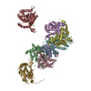
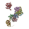
 Citation Citation | Journal: Proc Natl Acad Sci U S A / Year: 2021 Title: Actinobacteria challenge the paradigm: A unique protein architecture for a well-known, central metabolic complex. Authors: Eduardo M Bruch / Pierre Vilela / Lu Yang / Alexandra Boyko / Norik Lexa-Sapart / Bertrand Raynal / Pedro M Alzari / Marco Bellinzoni /  Abstract: α-oxoacid dehydrogenase complexes are large, tripartite enzymatic machineries carrying out key reactions in central metabolism. Extremely conserved across the tree of life, they have been, so far, ...α-oxoacid dehydrogenase complexes are large, tripartite enzymatic machineries carrying out key reactions in central metabolism. Extremely conserved across the tree of life, they have been, so far, all considered to be structured around a high-molecular weight hollow core, consisting of up to 60 subunits of the acyltransferase component. We provide here evidence that Actinobacteria break the rule by possessing an acetyltranferase component reduced to its minimally active, trimeric unit, characterized by a unique C-terminal helix bearing an actinobacterial specific insertion that precludes larger protein oligomerization. This particular feature, together with the presence of an gene coding for both the decarboxylase and the acyltransferase domains on the same polypetide, is spread over Actinobacteria and reflects the association of PDH and ODH into a single physical complex. Considering the central role of the pyruvate and 2-oxoglutarate nodes in central metabolism, our findings pave the way to both therapeutic and metabolic engineering applications. | ||||||
| History |
|
- Structure visualization
| Structure viewer | Molecule: MolmilJmol/JSmol |
|---|
- Downloads & links
-Download
| PDBx/mmCIF format | 6zzn.cif.gz | 106.1 KB | Display | PDBx/mmCIF format |
|---|---|---|---|---|
| PDB format | pdb6zzn.ent.gz | 82 KB | Display | PDB format |
| PDBx/mmJSON format | 6zzn.json.gz | Tree view | PDBx/mmJSON format | |
| Others |  Other downloads Other downloads |
-Validation report
| Arichive directory | https://data.pdbj.org/pub/pdb/validation_reports/zz/6zznftp://data.pdbj.org/pub/pdb/validation_reports/zz/6zzn | HTTPS FTP |
|---|
-Related structure data
| Related structure data | 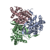 6zziC 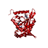 6zzjC 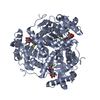 6zzkC 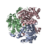 6zzlC  6zzmC 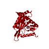 3l60S S: Starting model for refinement C: citing same article ( |
|---|---|
| Similar structure data |


-Links
 PDBj
PDBj


- Assembly
| Deposited unit | 
| |||||||||||||||
|---|---|---|---|---|---|---|---|---|---|---|---|---|---|---|---|---|
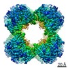
| 1 | x 24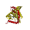
| |||||||||||||||
| Unit cell |
| |||||||||||||||
| Components on special symmetry positions |
|
-Components
| #1: Protein | Mass: 24704.479 Da / Num. of mol.: 1 Source method: isolated from a genetically manipulated source Source: (gene. exp.) Mycobacterium tuberculosis H37Rv (bacteria)Gene: bkdC, pdhC, Rv2495c / Production host: References: UniProt: O06159, dihydrolipoyllysine-residue (2-methylpropanoyl)transferase | ||||||
|---|---|---|---|---|---|---|---|
| #2: Chemical |   Mass: 59.044 Da / Num. of mol.: 2 / Source method: obtained synthetically / Formula: C2H3O2 Mass: 59.044 Da / Num. of mol.: 2 / Source method: obtained synthetically / Formula: C2H3O2#3: Chemical | ChemComp-IMD / |   Mass: 69.085 Da / Num. of mol.: 1 / Source method: obtained synthetically / Formula: C3H5N2 Mass: 69.085 Da / Num. of mol.: 1 / Source method: obtained synthetically / Formula: C3H5N2#4: Water | ChemComp-HOH / |  Mass: 18.015 Da / Num. of mol.: 337 / Source method: isolated from a natural source / Formula: H2O Mass: 18.015 Da / Num. of mol.: 337 / Source method: isolated from a natural source / Formula: H2OHas ligand of interest | N | |
-Experimental details
-Experiment
| Experiment | Method: X-RAY DIFFRACTION / Number of used crystals: 1 |
|---|
- Sample preparation
| Crystal grow | Temperature: 291 K / Method: vapor diffusion, sitting drop / pH: 6.5 / Details: 0.1 M imidazole pH 6.5, 4 M ammonium acetate |
|---|
-Data collection
| Diffraction | Mean temperature: 100 K / Serial crystal experiment: N |
|---|---|
| Diffraction source | Source: SYNCHROTRON / Site: ESRF / Beamline: MASSIF-1 / Wavelength: 0.966 Å |
| Detector | Type: DECTRIS PILATUS3 2M / Detector: PIXEL / Date: May 8, 2018 |
| Radiation | Protocol: SINGLE WAVELENGTH / Monochromatic (M) / Laue (L): M / Scattering type: x-ray |
| Radiation wavelength | Wavelength: 0.966 Å / Relative weight: 1 |
| Reflection | Resolution: 1.5→49.112 Å / Num. obs: 67274 / % possible obs: 100 % / Redundancy: 17.2 % / CC1/2: 0.999 / Rpim(I) all: 0.023 / Net I/σ(I): 15.2 |
| Reflection shell | Resolution: 1.5→1.526 Å / Num. unique obs: 3308 / CC1/2: 0.748 / Rpim(I) all: 0.391 |
- Processing
| Software |
| |||||||||||||||||||||||||||||||||||||||||||||||||||||||||||||||||||||||||||
|---|---|---|---|---|---|---|---|---|---|---|---|---|---|---|---|---|---|---|---|---|---|---|---|---|---|---|---|---|---|---|---|---|---|---|---|---|---|---|---|---|---|---|---|---|---|---|---|---|---|---|---|---|---|---|---|---|---|---|---|---|---|---|---|---|---|---|---|---|---|---|---|---|---|---|---|---|
| Refinement | Method to determine structure: MOLECULAR REPLACEMENT Starting model: 3L60 Resolution: 1.5→49.11 Å / Cor.coef. Fo:Fc: 0.951 / Cor.coef. Fo:Fc free: 0.961 / SU R Cruickshank DPI: 0.053 / Cross valid method: THROUGHOUT / SU R Blow DPI: 0.058 / SU Rfree Blow DPI: 0.055 / SU Rfree Cruickshank DPI: 0.05
| |||||||||||||||||||||||||||||||||||||||||||||||||||||||||||||||||||||||||||
| Displacement parameters | Biso mean: 28.74 Å2
| |||||||||||||||||||||||||||||||||||||||||||||||||||||||||||||||||||||||||||
| Refine analyze | Luzzati coordinate error obs: 0.19 Å | |||||||||||||||||||||||||||||||||||||||||||||||||||||||||||||||||||||||||||
| Refinement step | Cycle: LAST / Resolution: 1.5→49.11 Å
| |||||||||||||||||||||||||||||||||||||||||||||||||||||||||||||||||||||||||||
| Refine LS restraints |
| |||||||||||||||||||||||||||||||||||||||||||||||||||||||||||||||||||||||||||
| LS refinement shell | Resolution: 1.5→1.51 Å
| |||||||||||||||||||||||||||||||||||||||||||||||||||||||||||||||||||||||||||
| Refinement TLS params. | Refine-ID: X-RAY DIFFRACTION
| |||||||||||||||||||||||||||||||||||||||||||||||||||||||||||||||||||||||||||
| Refinement TLS group |
|
