 Movie
Movie Controller


+ Open data

- Basic information
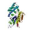
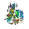
| Entry | Database: PDB / ID: 6zz9 | ||||||
|---|---|---|---|---|---|---|---|
| Title | Crystal structure of CbpB from Streptococcus agalactiae | ||||||
 Components Components | CBS domain-containing protein | ||||||
 Keywords Keywords | SIGNALING PROTEIN / CBS / c-di-AMP | ||||||
| Function / homology | : / CBS domain superfamily / CBS domain / CBS domain / CBS domain profile. / : / Cyclic-di-AMP-binding protein CbpB Function and homology information Function and homology information | ||||||
| Biological species |  Streptococcus agalactiae (bacteria) Streptococcus agalactiae (bacteria) | ||||||
| Method |  X-RAY DIFFRACTION / SYNCHROTRON / SAD / Resolution: 2.644 Å X-RAY DIFFRACTION / SYNCHROTRON / SAD / Resolution: 2.644 Å | ||||||
 Authors Authors | Mechaly, A.E. / Covaleda-Cortes, G. / Kaminski, P.A. | ||||||
 Citation Citation | Journal: Febs J. / Year: 2023 Title: The c-di-AMP-binding protein CbpB modulates the level of ppGpp alarmone in Streptococcus agalactiae. Authors: Covaleda-Cortes, G. / Mechaly, A. / Brissac, T. / Baehre, H. / Devaux, L. / England, P. / Raynal, B. / Hoos, S. / Gominet, M. / Firon, A. / Trieu-Cuot, P. / Kaminski, P.A. | ||||||
| History |
|
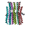
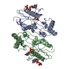
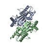
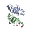
- Structure visualization
| Structure viewer | Molecule: MolmilJmol/JSmol |
|---|
- Downloads & links
-Download
| PDBx/mmCIF format | 6zz9.cif.gz | 138.2 KB | Display | PDBx/mmCIF format |
|---|---|---|---|---|
| PDB format | pdb6zz9.ent.gz | 110.5 KB | Display | PDB format |
| PDBx/mmJSON format | 6zz9.json.gz | Tree view | PDBx/mmJSON format | |
| Others |  Other downloads Other downloads |
-Validation report
| Arichive directory | https://data.pdbj.org/pub/pdb/validation_reports/zz/6zz9ftp://data.pdbj.org/pub/pdb/validation_reports/zz/6zz9 | HTTPS FTP |
|---|
-Related structure data


-Links
 PDBj
PDBj

- Assembly
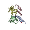
| Deposited unit | 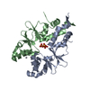
| ||||||||
|---|---|---|---|---|---|---|---|---|---|
| 1 |
| ||||||||
| Unit cell |
|
-Components
| #1: Protein | Mass: 19848.572 Da / Num. of mol.: 2 Source method: isolated from a genetically manipulated source Source: (gene. exp.) Streptococcus agalactiae (bacteria)Gene: ykuL, C6N06_01440, D5F95_08070, DX05_08405, E8E04_03435, F5F86_02645, NCTC9828_00932, WA02_02795, WA05_04310 Production host: #2: Chemical |   Mass: 96.063 Da / Num. of mol.: 3 / Source method: obtained synthetically / Formula: SO4 Mass: 96.063 Da / Num. of mol.: 3 / Source method: obtained synthetically / Formula: SO4#3: Chemical |   Mass: 195.078 Da / Num. of mol.: 2 / Source method: obtained synthetically / Formula: Pt Mass: 195.078 Da / Num. of mol.: 2 / Source method: obtained synthetically / Formula: PtHas ligand of interest | N | |
|---|
-Experimental details
-Experiment
| Experiment | Method: X-RAY DIFFRACTION / Number of used crystals: 1 |
|---|
- Sample preparation
| Crystal | Density Matthews: 2.2 Å3/Da / Density % sol: 44.17 % |
|---|---|
| Crystal grow | Temperature: 291 K / Method: vapor diffusion Details: 200 mM ammonium sulfate, 100 mM MES pH 6.5, 30% w/v PEG MME 5000 |
-Data collection
| Diffraction | Mean temperature: 100 K / Serial crystal experiment: N |
|---|---|
| Diffraction source | Source: SYNCHROTRON / Site: SOLEIL  / Beamline: PROXIMA 1 / Wavelength: 1.0716 Å / Beamline: PROXIMA 1 / Wavelength: 1.0716 Å |
| Detector | Type: DECTRIS PILATUS3 6M / Detector: PIXEL / Date: Jan 28, 2017 |
| Radiation | Protocol: SINGLE WAVELENGTH / Monochromatic (M) / Laue (L): M / Scattering type: x-ray |
| Radiation wavelength | Wavelength: 1.0716 Å / Relative weight: 1 |
| Reflection | Resolution: 2.644→47.62 Å / Num. obs: 11037 / % possible obs: 98.94 % / Redundancy: 4.3 % / CC1/2: 0.996 / CC star: 0.999 / Rmerge(I) obs: 0.06712 / Rpim(I) all: 0.03644 / Rrim(I) all: 0.07653 / Net I/σ(I): 10.76 |
| Reflection shell | Resolution: 2.644→2.739 Å / Redundancy: 4.2 % / Rmerge(I) obs: 1.346 / Num. unique obs: 1030 / CC1/2: 0.709 / CC star: 0.911 / Rpim(I) all: 0.7379 / Rrim(I) all: 1.538 / % possible all: 96.34 |
- Processing
| Software |
| |||||||||||||||||||||||||||||||||||||||||||||||||||||||||||||||||||||||||||
|---|---|---|---|---|---|---|---|---|---|---|---|---|---|---|---|---|---|---|---|---|---|---|---|---|---|---|---|---|---|---|---|---|---|---|---|---|---|---|---|---|---|---|---|---|---|---|---|---|---|---|---|---|---|---|---|---|---|---|---|---|---|---|---|---|---|---|---|---|---|---|---|---|---|---|---|---|
| Refinement | Method to determine structure: SAD / Resolution: 2.644→47.62 Å / Cor.coef. Fo:Fc: 0.938 / Cor.coef. Fo:Fc free: 0.932 / SU R Cruickshank DPI: 1.075 / Cross valid method: THROUGHOUT / SU R Blow DPI: 1.586 / SU Rfree Blow DPI: 0.322 / SU Rfree Cruickshank DPI: 0.321
| |||||||||||||||||||||||||||||||||||||||||||||||||||||||||||||||||||||||||||
| Displacement parameters | Biso mean: 100.59 Å2
| |||||||||||||||||||||||||||||||||||||||||||||||||||||||||||||||||||||||||||
| Refine analyze | Luzzati coordinate error obs: 0.46 Å | |||||||||||||||||||||||||||||||||||||||||||||||||||||||||||||||||||||||||||
| Refinement step | Cycle: LAST / Resolution: 2.644→47.62 Å
| |||||||||||||||||||||||||||||||||||||||||||||||||||||||||||||||||||||||||||
| Refine LS restraints |
| |||||||||||||||||||||||||||||||||||||||||||||||||||||||||||||||||||||||||||
| LS refinement shell | Resolution: 2.644→2.68 Å
| |||||||||||||||||||||||||||||||||||||||||||||||||||||||||||||||||||||||||||
| Refinement TLS params. | Refine-ID: X-RAY DIFFRACTION
| |||||||||||||||||||||||||||||||||||||||||||||||||||||||||||||||||||||||||||
| Refinement TLS group |
|
