 Movie
Movie Controller


+ Open data

- Basic information
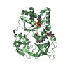
| Entry | Database: PDB / ID: 6zza | ||||||
|---|---|---|---|---|---|---|---|
| Title | Crystal structure of CbpB in complex with c-di-AMP | ||||||
 Components Components | CBS domain-containing protein | ||||||
 Keywords Keywords | SIGNALING PROTEIN / CBS / c-di-AMP | ||||||
| Function / homology | : / CBS domain superfamily / CBS domain / CBS domain / CBS domain profile. / Chem-2BA / Cyclic-di-AMP-binding protein CbpB Function and homology information Function and homology information | ||||||
| Biological species |  Streptococcus agalactiae (bacteria) Streptococcus agalactiae (bacteria) | ||||||
| Method |  X-RAY DIFFRACTION / SYNCHROTRON / MOLECULAR REPLACEMENT / Resolution: 2.491 Å X-RAY DIFFRACTION / SYNCHROTRON / MOLECULAR REPLACEMENT / Resolution: 2.491 Å | ||||||
 Authors Authors | Mechaly, A.E. / Covaleda-Cortes, G. / Kaminski, P.A. | ||||||
 Citation Citation | Journal: Febs J. / Year: 2023 Title: The c-di-AMP-binding protein CbpB modulates the level of ppGpp alarmone in Streptococcus agalactiae. Authors: Covaleda-Cortes, G. / Mechaly, A. / Brissac, T. / Baehre, H. / Devaux, L. / England, P. / Raynal, B. / Hoos, S. / Gominet, M. / Firon, A. / Trieu-Cuot, P. / Kaminski, P.A. | ||||||
| History |
|
- Structure visualization
| Structure viewer | Molecule: MolmilJmol/JSmol |
|---|
- Downloads & links
-Download
| PDBx/mmCIF format | 6zza.cif.gz | 82.6 KB | Display | PDBx/mmCIF format |
|---|---|---|---|---|
| PDB format | pdb6zza.ent.gz | 57.9 KB | Display | PDB format |
| PDBx/mmJSON format | 6zza.json.gz | Tree view | PDBx/mmJSON format | |
| Others |  Other downloads Other downloads |
-Validation report
| Arichive directory | https://data.pdbj.org/pub/pdb/validation_reports/zz/6zzaftp://data.pdbj.org/pub/pdb/validation_reports/zz/6zza | HTTPS FTP |
|---|
-Related structure data
| Related structure data | 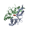 6zz9SC S: Starting model for refinement C: citing same article ( |
|---|---|
| Similar structure data |


-Links
 PDBj
PDBj
- Assembly
| Deposited unit | 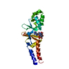
| ||||||||||||
|---|---|---|---|---|---|---|---|---|---|---|---|---|---|
| 1 | 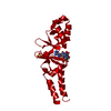
| ||||||||||||
| Unit cell |
|
-Components
| #1: Protein | Mass: 19848.572 Da / Num. of mol.: 1 Source method: isolated from a genetically manipulated source Source: (gene. exp.) Streptococcus agalactiae (bacteria)Gene: ykuL, C6N06_01440, D5F95_08070, DX05_08405, E8E04_03435, F5F86_02645, NCTC9828_00932, WA02_02795, WA05_04310 Production host: |
|---|---|
| #2: Chemical | ChemComp-2BA / (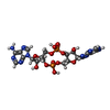  Mass: 658.412 Da / Num. of mol.: 1 / Source method: obtained synthetically / Formula: C20H24N10O12P2 / Feature type: SUBJECT OF INVESTIGATION Mass: 658.412 Da / Num. of mol.: 1 / Source method: obtained synthetically / Formula: C20H24N10O12P2 / Feature type: SUBJECT OF INVESTIGATION |
| #3: Chemical | ChemComp-SO4 /   Mass: 96.063 Da / Num. of mol.: 1 / Source method: obtained synthetically / Formula: SO4 Mass: 96.063 Da / Num. of mol.: 1 / Source method: obtained synthetically / Formula: SO4 |
| #4: Water | ChemComp-HOH /  Mass: 18.015 Da / Num. of mol.: 48 / Source method: isolated from a natural source / Formula: H2O Mass: 18.015 Da / Num. of mol.: 48 / Source method: isolated from a natural source / Formula: H2O |
| Has ligand of interest | Y |
| Has protein modification | N |
-Experimental details
-Experiment
| Experiment | Method: X-RAY DIFFRACTION / Number of used crystals: 1 |
|---|
- Sample preparation
| Crystal | Density Matthews: 2.15 Å3/Da / Density % sol: 42.82 % |
|---|---|
| Crystal grow | Temperature: 291 K / Method: vapor diffusion / Details: 200 mM ammonium sulfate, 20% w/v PEG 4000 |
-Data collection
| Diffraction | Mean temperature: 100 K / Serial crystal experiment: N |
|---|---|
| Diffraction source | Source: SYNCHROTRON / Site: SOLEIL  / Beamline: PROXIMA 2 / Wavelength: 0.9801 Å / Beamline: PROXIMA 2 / Wavelength: 0.9801 Å |
| Detector | Type: DECTRIS EIGER2 X 9M / Detector: PIXEL / Date: Jun 9, 2017 |
| Radiation | Protocol: SINGLE WAVELENGTH / Monochromatic (M) / Laue (L): M / Scattering type: x-ray |
| Radiation wavelength | Wavelength: 0.9801 Å / Relative weight: 1 |
| Reflection | Resolution: 2.491→31.05 Å / Num. obs: 6110 / % possible obs: 99.12 % / Redundancy: 5.3 % / CC1/2: 0.996 / CC star: 0.999 / Rmerge(I) obs: 0.1012 / Rpim(I) all: 0.04755 / Rrim(I) all: 0.1124 / Net I/σ(I): 9.79 |
| Reflection shell | Resolution: 2.491→2.58 Å / Redundancy: 5.1 % / Rmerge(I) obs: 0.6201 / Mean I/σ(I) obs: 2.03 / Num. unique obs: 578 / CC1/2: 0.798 / CC star: 0.942 / Rpim(I) all: 0.2914 / Rrim(I) all: 0.6887 / % possible all: 93.21 |
- Processing
| Software |
| ||||||||||||||||||||||||
|---|---|---|---|---|---|---|---|---|---|---|---|---|---|---|---|---|---|---|---|---|---|---|---|---|---|
| Refinement | Method to determine structure: MOLECULAR REPLACEMENT Starting model: 6ZZ9 Resolution: 2.491→31.05 Å / Cross valid method: THROUGHOUT Stereochemistry target values: GeoStd + Monomer Library + CDL v1.2
| ||||||||||||||||||||||||
| Displacement parameters | Biso mean: 67.54 Å2 | ||||||||||||||||||||||||
| Refinement step | Cycle: LAST / Resolution: 2.491→31.05 Å
| ||||||||||||||||||||||||
| Refine LS restraints |
| ||||||||||||||||||||||||
| LS refinement shell | Resolution: 2.491→2.78 Å
|
