 Movie
Movie Controller

[English] 日本語
 Yorodumi
- PDB-6zxj: Fully-loaded anthrax lethal toxin in its heptameric pre-pore stat... -
+ Open data

- Basic information
| Entry | Database: PDB / ID: 6zxj | ||||||
|---|---|---|---|---|---|---|---|
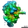
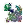
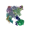
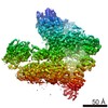
| Title | Fully-loaded anthrax lethal toxin in its heptameric pre-pore state, in which the third lethal factor is masked out (PA7LF3-masked) | ||||||
 Components Components |
| ||||||
 Keywords Keywords | TOXIN / anthrax lethal toxin / fully-loaded pre-pore state / membrane translocase / cytotoxic substrate | ||||||
| Function / homology |  Function and homology information Function and homology informationanthrax lethal factor endopeptidase / symbiont-mediated suppression of host MAPK cascade / host cell cytosol / Uptake and function of anthrax toxins / host cell endosome membrane / metalloendopeptidase activity / protein homooligomerization / metallopeptidase activity / toxin activity / host cell plasma membrane ...anthrax lethal factor endopeptidase / symbiont-mediated suppression of host MAPK cascade / host cell cytosol / Uptake and function of anthrax toxins / host cell endosome membrane / metalloendopeptidase activity / protein homooligomerization / metallopeptidase activity / toxin activity / host cell plasma membrane / proteolysis / extracellular region / zinc ion binding / metal ion binding / identical protein binding Similarity search - Function | ||||||
| Biological species |  | ||||||
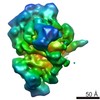
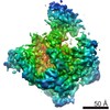
| Method | ELECTRON MICROSCOPY / single particle reconstruction / cryo EM / Resolution: 3.5 Å | ||||||
 Authors Authors | Quentin, D. / Antoni, C. / Gatsogiannis, C. / Raunser, S. | ||||||
| Funding support |  Germany, 1items Germany, 1items
| ||||||
 Citation Citation | Journal: PLoS Pathog / Year: 2020 Title: Cryo-EM structure of the fully-loaded asymmetric anthrax lethal toxin in its heptameric pre-pore state. Authors: Claudia Antoni / Dennis Quentin / Alexander E Lang / Klaus Aktories / Christos Gatsogiannis / Stefan Raunser / Abstract: Anthrax toxin is the major virulence factor secreted by Bacillus anthracis, causing high mortality in humans and other mammals. It consists of a membrane translocase, known as protective antigen (PA) ...Anthrax toxin is the major virulence factor secreted by Bacillus anthracis, causing high mortality in humans and other mammals. It consists of a membrane translocase, known as protective antigen (PA), that catalyzes the unfolding of its cytotoxic substrates lethal factor (LF) and edema factor (EF), followed by translocation into the host cell. Substrate recruitment to the heptameric PA pre-pore and subsequent translocation, however, are not well understood. Here, we report three high-resolution cryo-EM structures of the fully-loaded anthrax lethal toxin in its heptameric pre-pore state, which differ in the position and conformation of LFs. The structures reveal that three LFs interact with the heptameric PA and upon binding change their conformation to form a continuous chain of head-to-tail interactions. As a result of the underlying symmetry mismatch, one LF binding site in PA remains unoccupied. Whereas one LF directly interacts with a part of PA called α-clamp, the others do not interact with this region, indicating an intermediate state between toxin assembly and translocation. Interestingly, the interaction of the N-terminal domain with the α-clamp correlates with a higher flexibility in the C-terminal domain of the protein. Based on our data, we propose a model for toxin assembly, in which the relative position of the N-terminal α-helices in the three LFs determines which factor is translocated first. | ||||||
| History |
|
- Structure visualization
| Movie |
Movie viewer |
|---|---|
| Structure viewer | Molecule: MolmilJmol/JSmol |
- Downloads & links
-Download
| PDBx/mmCIF format | 6zxj.cif.gz | 870.3 KB | Display | PDBx/mmCIF format |
|---|---|---|---|---|
| PDB format | pdb6zxj.ent.gz | 699.4 KB | Display | PDB format |
| PDBx/mmJSON format | 6zxj.json.gz | Tree view | PDBx/mmJSON format | |
| Others |  Other downloads Other downloads |
-Validation report
| Arichive directory | https://data.pdbj.org/pub/pdb/validation_reports/zx/6zxjftp://data.pdbj.org/pub/pdb/validation_reports/zx/6zxj | HTTPS FTP |
|---|
-Related structure data
| Related structure data |  11522MC  6zxkC  6zxlC M: map data used to model this data C: citing same article ( |
|---|---|
| Similar structure data |


-Links
 PDBj
PDBj


- Assembly
| Deposited unit | 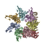
|
|---|---|
| 1 |
|
-Components
| #1: Protein | Mass: 85679.930 Da / Num. of mol.: 7 Source method: isolated from a genetically manipulated source Source: (gene. exp.) #2: Protein | Mass: 93904.211 Da / Num. of mol.: 2 Source method: isolated from a genetically manipulated source Source: (gene. exp.) References: UniProt: P15917, anthrax lethal factor endopeptidase |
|---|
-Experimental details
-Experiment
| Experiment | Method: ELECTRON MICROSCOPY |
|---|---|
| EM experiment | Aggregation state: PARTICLE / 3D reconstruction method: single particle reconstruction |
- Sample preparation
| Component |
| ||||||||||||||||||||||||||||
|---|---|---|---|---|---|---|---|---|---|---|---|---|---|---|---|---|---|---|---|---|---|---|---|---|---|---|---|---|---|
| Molecular weight |
| ||||||||||||||||||||||||||||
| Source (natural) |
| ||||||||||||||||||||||||||||
| Source (recombinant) |
| ||||||||||||||||||||||||||||
| Buffer solution | pH: 8.5 | ||||||||||||||||||||||||||||
| Buffer component |
| ||||||||||||||||||||||||||||
| Specimen | Conc.: 0.06 mg/ml / Embedding applied: NO / Shadowing applied: NO / Staining applied: NO / Vitrification applied: YES | ||||||||||||||||||||||||||||
| Specimen support | Grid material: COPPER / Grid mesh size: 300 divisions/in. / Grid type: Quantifoil R1.2/1.3 | ||||||||||||||||||||||||||||
| Vitrification | Instrument: GATAN CRYOPLUNGE 3 / Cryogen name: ETHANE / Humidity: 95 % / Chamber temperature: 286 K Details: 4 uL sample was applied to grid (with 2 nm additional carbon layer) and incubated for 45 s prior blotting. |
- Electron microscopy imaging
| Experimental equipment |  Model: Titan Krios / Image courtesy: FEI Company |
|---|---|
| Microscopy | Model: FEI TITAN KRIOS |
| Electron gun | Electron source:  FIELD EMISSION GUN / Accelerating voltage: 300 kV / Illumination mode: FLOOD BEAM FIELD EMISSION GUN / Accelerating voltage: 300 kV / Illumination mode: FLOOD BEAM |
| Electron lens | Mode: BRIGHT FIELD / Nominal magnification: 130000 X / Calibrated defocus min: 1200 nm / Calibrated defocus max: 2600 nm / Cs: 2.7 mm |
| Specimen holder | Cryogen: NITROGEN / Specimen holder model: FEI TITAN KRIOS AUTOGRID HOLDER |
| Image recording | Average exposure time: 15 sec. / Electron dose: 74.4 e/Å2 / Detector mode: COUNTING / Film or detector model: GATAN K2 SUMMIT (4k x 4k) / Num. of real images: 5238 |
| EM imaging optics | Energyfilter name: GIF Bioquantum / Energyfilter slit width: 20 eV |
| Image scans | Movie frames/image: 40 |
- Processing
| Software | Name: PHENIX / Version: 1.18.2_3874: / Classification: refinement | ||||||||||||||||||||||||||||||||||||||||
|---|---|---|---|---|---|---|---|---|---|---|---|---|---|---|---|---|---|---|---|---|---|---|---|---|---|---|---|---|---|---|---|---|---|---|---|---|---|---|---|---|---|
| EM software |
| ||||||||||||||||||||||||||||||||||||||||
| CTF correction | Type: PHASE FLIPPING AND AMPLITUDE CORRECTION | ||||||||||||||||||||||||||||||||||||||||
| Particle selection | Num. of particles selected: 382000 | ||||||||||||||||||||||||||||||||||||||||
| Symmetry | Point symmetry: C1 (asymmetric) | ||||||||||||||||||||||||||||||||||||||||
| 3D reconstruction | Resolution: 3.5 Å / Resolution method: FSC 0.143 CUT-OFF / Num. of particles: 210000 / Algorithm: BACK PROJECTION / Symmetry type: POINT | ||||||||||||||||||||||||||||||||||||||||
| Atomic model building | Protocol: FLEXIBLE FIT Details: The protective antigen (PA) monomer from 3HVD was placed seven times into the density corresponding to the heptameric PA ring using the rigid-body fit in Chimera. Two copies of the monomeric ...Details: The protective antigen (PA) monomer from 3HVD was placed seven times into the density corresponding to the heptameric PA ring using the rigid-body fit in Chimera. Two copies of the monomeric lethal factor (LF) from 1J7N were fitted similarly into the corresponding LF density located atop of the PA7 ring. The resulting model was then flexibly fitted using iMODFIT. The model was further refined in iterative rounds of phenix and coot. Unresolved regions were deleted and side chain information was removed for less well-defined regions. | ||||||||||||||||||||||||||||||||||||||||
| Atomic model building | 3D fitting-ID: 1 / Pdb chain-ID: A / Source name: PDB / Type: experimental model
| ||||||||||||||||||||||||||||||||||||||||
| Refine LS restraints |
|


