 Movie
Movie Controller

[English] 日本語
 Yorodumi
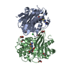
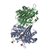
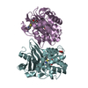
- PDB-6zw2: Crystal structure of OXA-10loop48 in complex with hydrolyzed meropenem -
+ Open data

- Basic information
| Entry | Database: PDB / ID: 6zw2 | ||||||
|---|---|---|---|---|---|---|---|
| Title | Crystal structure of OXA-10loop48 in complex with hydrolyzed meropenem | ||||||
 Components Components | Beta-lactamase | ||||||
 Keywords Keywords | HYDROLASE / Beta-lactamase / OXA-10loop48 / carbapenem / meropenem | ||||||
| Function / homology |  Function and homology information Function and homology informationpenicillin binding / antibiotic catabolic process / cell wall organization / beta-lactamase activity / beta-lactamase / response to antibiotic / metal ion binding / plasma membrane Similarity search - Function | ||||||
| Biological species |  | ||||||
| Method |  X-RAY DIFFRACTION / SYNCHROTRON / MOLECULAR REPLACEMENT / Resolution: 1.75 Å X-RAY DIFFRACTION / SYNCHROTRON / MOLECULAR REPLACEMENT / Resolution: 1.75 Å | ||||||
 Authors Authors | Tassone, G. / Di Pisa, F. / Benvenuti, M. / De Luca, F. / Pozzi, C. / Mangani, S. / Docquier, J.D. | ||||||
 Citation Citation | Journal: To Be Published Title: Mechanistic insights into carbapenem hydrolysis by OXA-48 and the OXA10-derived hybrids OXA-10 loop24 and loop48 Authors: Tassone, G. / Di Pisa, F. / Benvenuti, M. / De Luca, F. / Pozzi, C. / Mangani, S. / Docquier, J.D. | ||||||
| History |
|
- Structure visualization
| Structure viewer | Molecule: MolmilJmol/JSmol |
|---|
- Downloads & links
-Download
| PDBx/mmCIF format | 6zw2.cif.gz | 127.7 KB | Display | PDBx/mmCIF format |
|---|---|---|---|---|
| PDB format | pdb6zw2.ent.gz | 96 KB | Display | PDB format |
| PDBx/mmJSON format | 6zw2.json.gz | Tree view | PDBx/mmJSON format | |
| Others |  Other downloads Other downloads |
-Validation report
| Arichive directory | https://data.pdbj.org/pub/pdb/validation_reports/zw/6zw2ftp://data.pdbj.org/pub/pdb/validation_reports/zw/6zw2 | HTTPS FTP |
|---|
-Related structure data
| Related structure data | 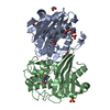 6zrgC  6zrhC  6zriC  6zrjC  6zrpC  3qncS C: citing same article ( S: Starting model for refinement |
|---|---|
| Similar structure data |


-Links
 PDBj
PDBj- Assembly
| Deposited unit | 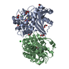
| ||||||||
|---|---|---|---|---|---|---|---|---|---|
| 1 |
| ||||||||
| Unit cell |
|
-Components
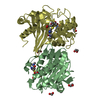
-Protein , 1 types, 2 molecules AB
| #1: Protein | Mass: 27568.412 Da / Num. of mol.: 2 Source method: isolated from a genetically manipulated source Source: (gene. exp.) Gene: blaoxa-10, blaOXA-10, oxa-10, BK373_28375, CQP61_30695, E4K55_27185, FORC82_p097, GII67_09965 Plasmid: pET24 / Production host: |
|---|
-Non-polymers , 5 types, 552 molecules 
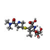


| #2: Chemical | ChemComp-EDO /  Mass: 62.068 Da / Num. of mol.: 8 / Source method: obtained synthetically / Formula: C2H6O2 Mass: 62.068 Da / Num. of mol.: 8 / Source method: obtained synthetically / Formula: C2H6O2#3: Chemical |  Mass: 401.478 Da / Num. of mol.: 2 / Source method: obtained synthetically / Formula: C17H27N3O6S Mass: 401.478 Da / Num. of mol.: 2 / Source method: obtained synthetically / Formula: C17H27N3O6SDetails: Other alternative conformations for hydrolysed meropenem are not excluded in the catalytic site for the observed positional disorder. Feature type: SUBJECT OF INVESTIGATION / Comment: antibiotic*YM #4: Chemical | ChemComp-SO4 / |  Mass: 96.063 Da / Num. of mol.: 1 / Source method: obtained synthetically / Formula: SO4 Mass: 96.063 Da / Num. of mol.: 1 / Source method: obtained synthetically / Formula: SO4#5: Chemical | ChemComp-CL /  Mass: 35.453 Da / Num. of mol.: 4 / Source method: obtained synthetically / Formula: Cl Mass: 35.453 Da / Num. of mol.: 4 / Source method: obtained synthetically / Formula: Cl#6: Water | ChemComp-HOH / | Mass: 18.015 Da / Num. of mol.: 537 / Source method: isolated from a natural source / Formula: H2O |
|---|
-Details
| Has ligand of interest | Y |
|---|
-Experimental details
-Experiment
| Experiment | Method: X-RAY DIFFRACTION / Number of used crystals: 1 |
|---|
- Sample preparation
| Crystal | Density Matthews: 2.68 Å3/Da / Density % sol: 54.16 % |
|---|---|
| Crystal grow | Temperature: 298 K / Method: vapor diffusion, sitting drop / pH: 9 / Details: 2.6 - 3 M ammonium sulfate and 100 mM bicine, pH 9 |
-Data collection
| Diffraction | Mean temperature: 100 K / Serial crystal experiment: N |
|---|---|
| Diffraction source | Source: SYNCHROTRON / Site: Diamond  / Beamline: I04-1 / Wavelength: 0.92 Å / Beamline: I04-1 / Wavelength: 0.92 Å |
| Detector | Type: DECTRIS PILATUS 6M-F / Detector: PIXEL / Date: Jul 5, 2013 |
| Radiation | Monochromator: Si(111) / Protocol: SINGLE WAVELENGTH / Monochromatic (M) / Laue (L): M / Scattering type: x-ray |
| Radiation wavelength | Wavelength: 0.92 Å / Relative weight: 1 |
| Reflection | Resolution: 1.75→52.45 Å / Num. obs: 60314 / % possible obs: 99.8 % / Redundancy: 6.6 % / Biso Wilson estimate: 16.1 Å2 / CC1/2: 0.997 / Rmerge(I) obs: 0.101 / Rpim(I) all: 0.042 / Rrim(I) all: 0.109 / Net I/σ(I): 11.5 |
| Reflection shell | Resolution: 1.75→1.84 Å / Redundancy: 6.1 % / Rmerge(I) obs: 0.713 / Mean I/σ(I) obs: 2.4 / Num. unique obs: 8684 / CC1/2: 0.807 / Rpim(I) all: 0.31 / Rrim(I) all: 0.78 / % possible all: 99.7 |
- Processing
| Software |
| |||||||||||||||||||||||||||||||||||||||||||||
|---|---|---|---|---|---|---|---|---|---|---|---|---|---|---|---|---|---|---|---|---|---|---|---|---|---|---|---|---|---|---|---|---|---|---|---|---|---|---|---|---|---|---|---|---|---|---|
| Refinement | Method to determine structure: MOLECULAR REPLACEMENT Starting model: 3QNC Resolution: 1.75→52.45 Å / Cor.coef. Fo:Fc: 0.959 / Cor.coef. Fo:Fc free: 0.938 / SU B: 2.622 / SU ML: 0.083 / Cross valid method: THROUGHOUT / σ(F): 0 / ESU R: 0.11 / ESU R Free: 0.11 / Stereochemistry target values: MAXIMUM LIKELIHOOD / Details: U VALUES : REFINED INDIVIDUALLY
| |||||||||||||||||||||||||||||||||||||||||||||
| Solvent computation | Ion probe radii: 0.8 Å / Shrinkage radii: 0.8 Å / VDW probe radii: 1.2 Å / Solvent model: MASK | |||||||||||||||||||||||||||||||||||||||||||||
| Displacement parameters | Biso max: 83.7 Å2 / Biso mean: 23.264 Å2 / Biso min: 7.71 Å2
| |||||||||||||||||||||||||||||||||||||||||||||
| Refine analyze | Luzzati coordinate error obs: 0.1935 Å | |||||||||||||||||||||||||||||||||||||||||||||
| Refinement step | Cycle: final / Resolution: 1.75→52.45 Å
| |||||||||||||||||||||||||||||||||||||||||||||
| Refine LS restraints |
| |||||||||||||||||||||||||||||||||||||||||||||
| LS refinement shell | Resolution: 1.75→1.795 Å / Rfactor Rfree error: 0 / Total num. of bins used: 20
|
