 Movie
Movie Controller

[English] 日本語
 Yorodumi
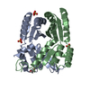
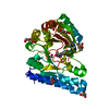
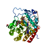
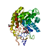
- PDB-6zv9: Terbium(III)-bound de novo TIM barrel-ferredoxin fold fusion dime... -
+ Open data

- Basic information
| Entry | Database: PDB / ID: 6zv9 | ||||||
|---|---|---|---|---|---|---|---|
| Title | Terbium(III)-bound de novo TIM barrel-ferredoxin fold fusion dimer with 4-glutamate binding site and tryptophan antenna (TFD-EE N6W) | ||||||
 Components Components | TFD-EE | ||||||
 Keywords Keywords | DE NOVO PROTEIN / Lanthanide-binding protein | ||||||
| Function / homology | Ribosomal protein S10 / Alpha-Beta Plaits / 2-Layer Sandwich / Alpha Beta / TERBIUM(III) ION Function and homology information Function and homology information | ||||||
| Biological species | synthetic construct (others) | ||||||
| Method |  X-RAY DIFFRACTION / SYNCHROTRON / MOLECULAR REPLACEMENT / Resolution: 1.85 Å X-RAY DIFFRACTION / SYNCHROTRON / MOLECULAR REPLACEMENT / Resolution: 1.85 Å | ||||||
 Authors Authors | Caldwell, S. / Haydon, I. / Piperidou, N. / Huang, P. / Hilvert, D. / Baker, D. / Zeymer, C. | ||||||
| Funding support |  Switzerland, 1items Switzerland, 1items
| ||||||
 Citation Citation | Journal: Proc.Natl.Acad.Sci.USA / Year: 2020 Title: Tight and specific lanthanide binding in a de novo TIM barrel with a large internal cavity designed by symmetric domain fusion. Authors: Caldwell, S.J. / Haydon, I.C. / Piperidou, N. / Huang, P.S. / Bick, M.J. / Sjostrom, H.S. / Hilvert, D. / Baker, D. / Zeymer, C. | ||||||
| History |
|
- Structure visualization
| Structure viewer | Molecule: MolmilJmol/JSmol |
|---|
- Downloads & links
-Download
| PDBx/mmCIF format | 6zv9.cif.gz | 145.7 KB | Display | PDBx/mmCIF format |
|---|---|---|---|---|
| PDB format | pdb6zv9.ent.gz | 114.7 KB | Display | PDB format |
| PDBx/mmJSON format | 6zv9.json.gz | Tree view | PDBx/mmJSON format | |
| Others |  Other downloads Other downloads |
-Validation report
| Arichive directory | https://data.pdbj.org/pub/pdb/validation_reports/zv/6zv9ftp://data.pdbj.org/pub/pdb/validation_reports/zv/6zv9 | HTTPS FTP |
|---|
-Related structure data


-Links
 PDBj
PDBj
- Assembly
| Deposited unit | 
| ||||||||
|---|---|---|---|---|---|---|---|---|---|
| 1 |
| ||||||||
| Unit cell |
|
-Components
| #1: Protein | Mass: 18906.947 Da / Num. of mol.: 2 / Mutation: N6W Source method: isolated from a genetically manipulated source Details: The first four amino acids (GAMG) are not part of the de novo designed sequence, but a results of the cloning and purification strategy. They are thus numbered -3, -2, -1, 0. Source: (gene. exp.) synthetic construct (others) / Production host:  #2: Chemical | ChemComp-TB / |   Mass: 158.925 Da / Num. of mol.: 1 / Source method: obtained synthetically / Formula: Tb / Feature type: SUBJECT OF INVESTIGATION Mass: 158.925 Da / Num. of mol.: 1 / Source method: obtained synthetically / Formula: Tb / Feature type: SUBJECT OF INVESTIGATION#3: Chemical | ChemComp-EDO / |   Mass: 62.068 Da / Num. of mol.: 1 / Source method: obtained synthetically / Formula: C2H6O2 Mass: 62.068 Da / Num. of mol.: 1 / Source method: obtained synthetically / Formula: C2H6O2#4: Water | ChemComp-HOH / |  Mass: 18.015 Da / Num. of mol.: 44 / Source method: isolated from a natural source / Formula: H2O Mass: 18.015 Da / Num. of mol.: 44 / Source method: isolated from a natural source / Formula: H2OHas ligand of interest | Y | |
|---|
-Experimental details
-Experiment
| Experiment | Method: X-RAY DIFFRACTION / Number of used crystals: 1 |
|---|
- Sample preparation
| Crystal | Density Matthews: 2.29 Å3/Da / Density % sol: 46.33 % |
|---|---|
| Crystal grow | Temperature: 293 K / Method: vapor diffusion Details: 0.1 M HEPES pH 7.5, 20 % PEG 4000, and 10 % iso-propanol, containing Gly3 as an additive |
-Data collection
| Diffraction | Mean temperature: 100 K / Serial crystal experiment: N |
|---|---|
| Diffraction source | Source: SYNCHROTRON / Site: SLS / Beamline: X06DA / Wavelength: 1 Å |
| Detector | Type: DECTRIS PILATUS 2M-F / Detector: PIXEL / Date: Jan 29, 2020 |
| Radiation | Protocol: SINGLE WAVELENGTH / Monochromatic (M) / Laue (L): M / Scattering type: x-ray |
| Radiation wavelength | Wavelength: 1 Å / Relative weight: 1 |
| Reflection | Resolution: 1.85→30 Å / Num. obs: 56427 / % possible obs: 98.4 % / Redundancy: 3.06 % / CC1/2: 0.999 / Rrim(I) all: 0.055 / Net I/σ(I): 13.12 |
| Reflection shell | Resolution: 1.85→1.95 Å / Redundancy: 2.97 % / Mean I/σ(I) obs: 1.94 / Num. unique obs: 8189 / CC1/2: 0.845 / Rrim(I) all: 0.697 / % possible all: 97.9 |
- Processing
| Software |
| |||||||||||||||||||||||||||||||||||||||||||||||||||||||||||||||||||||||||||
|---|---|---|---|---|---|---|---|---|---|---|---|---|---|---|---|---|---|---|---|---|---|---|---|---|---|---|---|---|---|---|---|---|---|---|---|---|---|---|---|---|---|---|---|---|---|---|---|---|---|---|---|---|---|---|---|---|---|---|---|---|---|---|---|---|---|---|---|---|---|---|---|---|---|---|---|---|
| Refinement | Method to determine structure: MOLECULAR REPLACEMENT Starting model: Single domain Rosetta models Resolution: 1.85→30 Å / Cor.coef. Fo:Fc: 0.962 / Cor.coef. Fo:Fc free: 0.952 / SU B: 9.792 / SU ML: 0.123 / Cross valid method: THROUGHOUT / σ(F): 0 / ESU R: 0.3 / ESU R Free: 0.135 / Stereochemistry target values: MAXIMUM LIKELIHOOD Details: HYDROGENS HAVE BEEN ADDED IN THE RIDING POSITIONS U VALUES : WITH TLS ADDED
| |||||||||||||||||||||||||||||||||||||||||||||||||||||||||||||||||||||||||||
| Solvent computation | Ion probe radii: 0.8 Å / Shrinkage radii: 0.8 Å / VDW probe radii: 1.2 Å / Solvent model: MASK | |||||||||||||||||||||||||||||||||||||||||||||||||||||||||||||||||||||||||||
| Displacement parameters | Biso max: 101.69 Å2 / Biso mean: 47.312 Å2 / Biso min: 22.11 Å2
| |||||||||||||||||||||||||||||||||||||||||||||||||||||||||||||||||||||||||||
| Refinement step | Cycle: final / Resolution: 1.85→30 Å
| |||||||||||||||||||||||||||||||||||||||||||||||||||||||||||||||||||||||||||
| Refine LS restraints |
| |||||||||||||||||||||||||||||||||||||||||||||||||||||||||||||||||||||||||||
| LS refinement shell | Resolution: 1.85→1.898 Å / Rfactor Rfree error: 0 / Total num. of bins used: 20
| |||||||||||||||||||||||||||||||||||||||||||||||||||||||||||||||||||||||||||
| Refinement TLS params. | Method: refined / Refine-ID: X-RAY DIFFRACTION
| |||||||||||||||||||||||||||||||||||||||||||||||||||||||||||||||||||||||||||
| Refinement TLS group |
|
