 Movie
Movie Controller


+ Open data

- Basic information
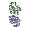
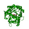
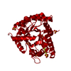
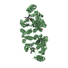
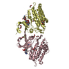
| Entry | Database: PDB / ID: 6vgo | ||||||
|---|---|---|---|---|---|---|---|
| Title | Crystal Structure of Human Dipeptidase 3 | ||||||
 Components Components | Dipeptidase 3 | ||||||
 Keywords Keywords | HYDROLASE / Metalloprotease | ||||||
| Function / homology |  Function and homology information Function and homology informationleukotriene D4 catabolic process / dipeptidase activity / side of membrane / acrosomal vesicle / proteolysis / membrane / plasma membrane Similarity search - Function | ||||||
| Biological species |  Homo sapiens (human) Homo sapiens (human) | ||||||
| Method |  X-RAY DIFFRACTION / SYNCHROTRON / MOLECULAR REPLACEMENT / Resolution: 1.82 Å X-RAY DIFFRACTION / SYNCHROTRON / MOLECULAR REPLACEMENT / Resolution: 1.82 Å | ||||||
 Authors Authors | Hayashi, K. / Longenecker, K.L. / Vivona, S. | ||||||
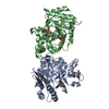
 Citation Citation | Journal: J.Struct.Biol. / Year: 2020 Title: Structure of human DPEP3 in complex with the SC-003 antibody Fab fragment reveals basis for lack of dipeptidase activity. Authors: Hayashi, K. / Longenecker, K.L. / Koenig, P. / Prashar, A. / Hampl, J. / Stoll, V. / Vivona, S. | ||||||
| History |
|
- Structure visualization
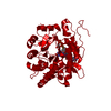
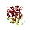
| Structure viewer | Molecule: MolmilJmol/JSmol |
|---|
- Downloads & links
-Download
| PDBx/mmCIF format | 6vgo.cif.gz | 88.9 KB | Display | PDBx/mmCIF format |
|---|---|---|---|---|
| PDB format | pdb6vgo.ent.gz | 64.3 KB | Display | PDB format |
| PDBx/mmJSON format | 6vgo.json.gz | Tree view | PDBx/mmJSON format | |
| Others |  Other downloads Other downloads |
-Validation report
| Arichive directory | https://data.pdbj.org/pub/pdb/validation_reports/vg/6vgoftp://data.pdbj.org/pub/pdb/validation_reports/vg/6vgo | HTTPS FTP |
|---|
-Related structure data
| Related structure data |  6vgrC  1itqS S: Starting model for refinement C: citing same article ( |
|---|---|
| Similar structure data |


-Links
 PDBj
PDBj

- Assembly
| Deposited unit | 
| ||||||||
|---|---|---|---|---|---|---|---|---|---|
| 1 | 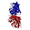
| ||||||||
| Unit cell |
|
-Components
| #1: Protein | Mass: 53745.145 Da / Num. of mol.: 1 Source method: isolated from a genetically manipulated source Source: (gene. exp.) Homo sapiens (human) / Gene: DPEP3, UNQ834/PRO1772 / Production host:   Cricetulus griseus (Chinese hamster) / References: UniProt: Q9H4B8, membrane dipeptidase Cricetulus griseus (Chinese hamster) / References: UniProt: Q9H4B8, membrane dipeptidase |
|---|---|
| #2: Water | ChemComp-HOH /  Mass: 18.015 Da / Num. of mol.: 220 / Source method: isolated from a natural source / Formula: H2O Mass: 18.015 Da / Num. of mol.: 220 / Source method: isolated from a natural source / Formula: H2O |
| Has protein modification | Y |
-Experimental details
-Experiment
| Experiment | Method: X-RAY DIFFRACTION / Number of used crystals: 1 |
|---|
- Sample preparation
| Crystal | Density Matthews: 1.96 Å3/Da / Density % sol: 37.29 % |
|---|---|
| Crystal grow | Temperature: 297 K / Method: vapor diffusion, sitting drop Details: 0.1 M calcium acetate, 0.1 M sodium acetate pH 4.5, 10% w/v PEG4000 |
-Data collection
| Diffraction | Mean temperature: 297 K / Serial crystal experiment: N |
|---|---|
| Diffraction source | Source: SYNCHROTRON / Site: APS  / Beamline: 17-ID / Wavelength: 1 Å / Beamline: 17-ID / Wavelength: 1 Å |
| Detector | Type: DECTRIS PILATUS 6M / Detector: PIXEL / Date: Dec 10, 2018 |
| Radiation | Protocol: SINGLE WAVELENGTH / Monochromatic (M) / Laue (L): M / Scattering type: x-ray |
| Radiation wavelength | Wavelength: 1 Å / Relative weight: 1 |
| Reflection | Resolution: 1.82→62.39 Å / Num. obs: 39548 / % possible obs: 99.8 % / Redundancy: 12.7 % / Rrim(I) all: 0.078 / Net I/σ(I): 18 |
| Reflection shell | Resolution: 1.82→1.85 Å / Num. unique obs: 1939 / Rpim(I) all: 0.38 |
- Processing
| Software |
| ||||||||||||||||||||||||||||||||||||||||||||||||||||||||||||||||||||||||||||||||||||||||||||||||||||||||||||
|---|---|---|---|---|---|---|---|---|---|---|---|---|---|---|---|---|---|---|---|---|---|---|---|---|---|---|---|---|---|---|---|---|---|---|---|---|---|---|---|---|---|---|---|---|---|---|---|---|---|---|---|---|---|---|---|---|---|---|---|---|---|---|---|---|---|---|---|---|---|---|---|---|---|---|---|---|---|---|---|---|---|---|---|---|---|---|---|---|---|---|---|---|---|---|---|---|---|---|---|---|---|---|---|---|---|---|---|---|---|
| Refinement | Method to determine structure: MOLECULAR REPLACEMENT Starting model: 1ITQ Resolution: 1.82→62.39 Å / Cor.coef. Fo:Fc: 0.949 / Cor.coef. Fo:Fc free: 0.941 / SU R Cruickshank DPI: 0.11 / Cross valid method: THROUGHOUT / σ(F): 0 / SU R Blow DPI: 0.117 / SU Rfree Blow DPI: 0.11 / SU Rfree Cruickshank DPI: 0.106
| ||||||||||||||||||||||||||||||||||||||||||||||||||||||||||||||||||||||||||||||||||||||||||||||||||||||||||||
| Displacement parameters | Biso max: 116.41 Å2 / Biso mean: 39.11 Å2 / Biso min: 24.14 Å2
| ||||||||||||||||||||||||||||||||||||||||||||||||||||||||||||||||||||||||||||||||||||||||||||||||||||||||||||
| Refine analyze | Luzzati coordinate error obs: 0.22 Å | ||||||||||||||||||||||||||||||||||||||||||||||||||||||||||||||||||||||||||||||||||||||||||||||||||||||||||||
| Refinement step | Cycle: final / Resolution: 1.82→62.39 Å
| ||||||||||||||||||||||||||||||||||||||||||||||||||||||||||||||||||||||||||||||||||||||||||||||||||||||||||||
| Refine LS restraints |
| ||||||||||||||||||||||||||||||||||||||||||||||||||||||||||||||||||||||||||||||||||||||||||||||||||||||||||||
| LS refinement shell | Resolution: 1.82→1.83 Å / Rfactor Rfree error: 0 / Total num. of bins used: 50
|
