 Movie
Movie Controller

[English] 日本語
 Yorodumi
- PDB-3gx9: Structure of morphinone reductase N189A mutant in complex with te... -
+ Open data

- Basic information
| Entry | Database: PDB / ID: 3gx9 | ||||||
|---|---|---|---|---|---|---|---|
| Title | Structure of morphinone reductase N189A mutant in complex with tetrahydroNAD | ||||||
 Components Components | Morphinone reductase | ||||||
 Keywords Keywords | OXIDOREDUCTASE / H-tunnelling / flavoprotein / NADH / morphinone reductase / hydride transfer | ||||||
| Function / homology |  Function and homology information Function and homology informationoxidoreductase activity, acting on the CH-CH group of donors, NAD or NADP as acceptor / FMN binding / cytosol Similarity search - Function | ||||||
| Biological species |  Pseudomonas putida (bacteria) Pseudomonas putida (bacteria) | ||||||
| Method |  X-RAY DIFFRACTION / SYNCHROTRON / MOLECULAR REPLACEMENT / Resolution: 2.28 Å X-RAY DIFFRACTION / SYNCHROTRON / MOLECULAR REPLACEMENT / Resolution: 2.28 Å | ||||||
 Authors Authors | Lafite, P. / Scrutton, N.S. / Leys, D. | ||||||
 Citation Citation | Journal: Chembiochem / Year: 2009 Title: Parallel Pathways and Free-Energy Landscapes for Enzymatic Hydride Transfer Probed by Hydrostatic Pressure Authors: Pudney, C.R. / McGrory, T. / Lafite, P. / Pang, J. / Hay, S. / Leys, D. / Sutcliffe, M.J. / Scrutton, N.S. | ||||||
| History |
|
- Structure visualization
| Structure viewer | Molecule: MolmilJmol/JSmol |
|---|
- Downloads & links
-Download
| PDBx/mmCIF format | 3gx9.cif.gz | 90.2 KB | Display | PDBx/mmCIF format |
|---|---|---|---|---|
| PDB format | pdb3gx9.ent.gz | 66.9 KB | Display | PDB format |
| PDBx/mmJSON format | 3gx9.json.gz | Tree view | PDBx/mmJSON format | |
| Others |  Other downloads Other downloads |
-Validation report
| Arichive directory | https://data.pdbj.org/pub/pdb/validation_reports/gx/3gx9ftp://data.pdbj.org/pub/pdb/validation_reports/gx/3gx9 | HTTPS FTP |
|---|
-Related structure data
| Related structure data |  2r14S S: Starting model for refinement |
|---|---|
| Similar structure data |


-Links
 PDBj
PDBj- Assembly
| Deposited unit | 
| ||||||||
|---|---|---|---|---|---|---|---|---|---|
| 1 | 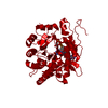
| ||||||||
| Unit cell |
| ||||||||
| Components on special symmetry positions |
|
-Components
| #1: Protein | Mass: 41247.867 Da / Num. of mol.: 1 / Mutation: N189A Source method: isolated from a genetically manipulated source Source: (gene. exp.) Pseudomonas putida (bacteria) / Strain: M10 / Gene: morB / Plasmid: pMORB2 / Production host: |
|---|---|
| #2: Chemical | ChemComp-FMN /   Mass: 456.344 Da / Num. of mol.: 1 / Source method: obtained synthetically / Formula: C17H21N4O9P Mass: 456.344 Da / Num. of mol.: 1 / Source method: obtained synthetically / Formula: C17H21N4O9P |
| #3: Chemical | ChemComp-TXD /   Mass: 667.457 Da / Num. of mol.: 1 / Source method: obtained synthetically / Formula: C21H31N7O14P2 Mass: 667.457 Da / Num. of mol.: 1 / Source method: obtained synthetically / Formula: C21H31N7O14P2 |
| #4: Water | ChemComp-HOH /  Mass: 18.015 Da / Num. of mol.: 174 / Source method: isolated from a natural source / Formula: H2O Mass: 18.015 Da / Num. of mol.: 174 / Source method: isolated from a natural source / Formula: H2O |
-Experimental details
-Experiment
| Experiment | Method: X-RAY DIFFRACTION / Number of used crystals: 1 |
|---|
- Sample preparation
| Crystal | Density Matthews: 3.32 Å3/Da / Density % sol: 62.97 % |
|---|---|
| Crystal grow | Temperature: 277 K / Method: vapor diffusion, sitting drop / pH: 7.5 Details: 0.1M Hepes, pH7.5, 0.1M MgCl2, 35% PEG400, Crystals were soaked in a 50% Ammonium sulfate Solution, containing 100mM NADH4, VAPOR DIFFUSION, SITTING DROP, temperature 277K |
-Data collection
| Diffraction | Mean temperature: 100 K |
|---|---|
| Diffraction source | Source: SYNCHROTRON / Site: Diamond  / Beamline: I04 / Wavelength: 0.9696 Å / Beamline: I04 / Wavelength: 0.9696 Å |
| Detector | Type: ADSC QUANTUM 315r / Detector: CCD / Date: Apr 18, 2008 |
| Radiation | Monochromator: Double Crystal Monochromator / Protocol: SINGLE WAVELENGTH / Monochromatic (M) / Laue (L): M / Scattering type: x-ray |
| Radiation wavelength | Wavelength: 0.9696 Å / Relative weight: 1 |
| Reflection | Resolution: 2.28→44.9 Å / Num. obs: 23517 / % possible obs: 92.1 % / Observed criterion σ(F): 0 / Observed criterion σ(I): 0 / Rmerge(I) obs: 0.077 / Net I/σ(I): 7.4 |
| Reflection shell | Resolution: 2.28→2.5 Å / Rmerge(I) obs: 0.291 / Mean I/σ(I) obs: 2.5 / % possible all: 94.6 |
- Processing
| Software |
| |||||||||||||||||||||||||||||||||||||||||||||||||||||||||||||||||
|---|---|---|---|---|---|---|---|---|---|---|---|---|---|---|---|---|---|---|---|---|---|---|---|---|---|---|---|---|---|---|---|---|---|---|---|---|---|---|---|---|---|---|---|---|---|---|---|---|---|---|---|---|---|---|---|---|---|---|---|---|---|---|---|---|---|---|
| Refinement | Method to determine structure: MOLECULAR REPLACEMENT Starting model: PDB ENTRY 2R14 Resolution: 2.28→44.9 Å / Cor.coef. Fo:Fc: 0.946 / Cor.coef. Fo:Fc free: 0.921 / SU B: 5.289 / SU ML: 0.129 / Cross valid method: THROUGHOUT / σ(F): 0 / σ(I): 0 / ESU R: 0.242 / ESU R Free: 0.203 / Stereochemistry target values: MAXIMUM LIKELIHOOD / Details: HYDROGENS HAVE BEEN ADDED IN THE RIDING POSITIONS
| |||||||||||||||||||||||||||||||||||||||||||||||||||||||||||||||||
| Solvent computation | Ion probe radii: 0.8 Å / Shrinkage radii: 0.8 Å / VDW probe radii: 1.2 Å / Solvent model: MASK | |||||||||||||||||||||||||||||||||||||||||||||||||||||||||||||||||
| Displacement parameters | Biso mean: 27.016 Å2
| |||||||||||||||||||||||||||||||||||||||||||||||||||||||||||||||||
| Refinement step | Cycle: LAST / Resolution: 2.28→44.9 Å
| |||||||||||||||||||||||||||||||||||||||||||||||||||||||||||||||||
| Refine LS restraints |
| |||||||||||||||||||||||||||||||||||||||||||||||||||||||||||||||||
| LS refinement shell | Resolution: 2.279→2.338 Å / Total num. of bins used: 20
|
