 Movie
Movie Controller

[English] 日本語
 Yorodumi
- PDB-6zsk: Cytochrome c prime beta from Methylococcus capsulatus (Bath): CO ... -
+ Open data

- Basic information
| Entry | Database: PDB / ID: 6zsk | ||||||
|---|---|---|---|---|---|---|---|
| Title | Cytochrome c prime beta from Methylococcus capsulatus (Bath): CO complex | ||||||
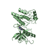
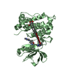
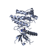
 Components Components | Cytochrome c | ||||||
 Keywords Keywords | OXIDOREDUCTASE / cytochrome c'-beta / methanotroph / carbon monoxide | ||||||
| Function / homology | Cytochrome P460 / Cytochrome P460 superfamily / Cytochrome P460 / metal ion binding / CARBON MONOXIDE / HEME C / Cytochrome c Function and homology information Function and homology information | ||||||
| Biological species |  Methylococcus capsulatus (bacteria) Methylococcus capsulatus (bacteria) | ||||||
| Method |  X-RAY DIFFRACTION / SYNCHROTRON / MOLECULAR REPLACEMENT / Resolution: 1.6 Å X-RAY DIFFRACTION / SYNCHROTRON / MOLECULAR REPLACEMENT / Resolution: 1.6 Å | ||||||
 Authors Authors | Adams, H.R. / Hough, M.A. | ||||||
 Citation Citation | Journal: To Be Published Title: to be published Authors: Adams, H.R. / Strange, R.W. / Svistunenko, D.A. / Andrew, C.R. / Hough, M.A. | ||||||
| History |
|
- Structure visualization
| Structure viewer | Molecule: MolmilJmol/JSmol |
|---|
- Downloads & links
-Download
| PDBx/mmCIF format | 6zsk.cif.gz | 76.2 KB | Display | PDBx/mmCIF format |
|---|---|---|---|---|
| PDB format | pdb6zsk.ent.gz | 55.2 KB | Display | PDB format |
| PDBx/mmJSON format | 6zsk.json.gz | Tree view | PDBx/mmJSON format | |
| Others |  Other downloads Other downloads |
-Validation report
| Summary document | 6zsk_validation.pdf.gz | 467.6 KB | Display | wwPDB validaton report |
|---|---|---|---|---|
| Full document | 6zsk_full_validation.pdf.gz | 469.2 KB | Display | |
| Data in XML | 6zsk_validation.xml.gz | 14.2 KB | Display | |
| Data in CIF | 6zsk_validation.cif.gz | 19.6 KB | Display | |
| Arichive directory | https://data.pdbj.org/pub/pdb/validation_reports/zs/6zskftp://data.pdbj.org/pub/pdb/validation_reports/zs/6zsk | HTTPS FTP |
-Related structure data
| Related structure data |  6hihS S: Starting model for refinement |
|---|---|
| Similar structure data |


-Links
 PDBj
PDBj


- Assembly
| Deposited unit | 
| ||||||||||||
|---|---|---|---|---|---|---|---|---|---|---|---|---|---|
| 1 |
| ||||||||||||
| Unit cell |
| ||||||||||||
| Components on special symmetry positions |
|
-Components
-Protein , 1 types, 2 molecules AB
| #1: Protein | Mass: 17370.650 Da / Num. of mol.: 2 Source method: isolated from a genetically manipulated source Source: (gene. exp.) Methylococcus capsulatus (strain ATCC 33009 / NCIMB 11132 / Bath) (bacteria)Strain: ATCC 33009 / NCIMB 11132 / Bath / Gene: ccp, MCA2394 / Production host: |
|---|
-Non-polymers , 5 types, 140 molecules 


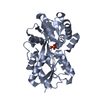
| #2: Chemical |  Mass: 618.503 Da / Num. of mol.: 2 / Source method: obtained synthetically / Formula: C34H34FeN4O4 Mass: 618.503 Da / Num. of mol.: 2 / Source method: obtained synthetically / Formula: C34H34FeN4O4#3: Chemical |  Mass: 65.409 Da / Num. of mol.: 3 / Source method: obtained synthetically / Formula: Zn Mass: 65.409 Da / Num. of mol.: 3 / Source method: obtained synthetically / Formula: Zn#4: Chemical |  Mass: 92.094 Da / Num. of mol.: 2 / Source method: obtained synthetically / Formula: C3H8O3 Mass: 92.094 Da / Num. of mol.: 2 / Source method: obtained synthetically / Formula: C3H8O3#5: Chemical |  Mass: 28.010 Da / Num. of mol.: 2 / Source method: obtained synthetically / Formula: CO / Feature type: SUBJECT OF INVESTIGATION Mass: 28.010 Da / Num. of mol.: 2 / Source method: obtained synthetically / Formula: CO / Feature type: SUBJECT OF INVESTIGATION#6: Water | ChemComp-HOH / | Mass: 18.015 Da / Num. of mol.: 131 / Source method: isolated from a natural source / Formula: H2O |
|---|
-Details
| Has ligand of interest | Y |
|---|---|
| Has protein modification | Y |
-Experimental details
-Experiment
| Experiment | Method: X-RAY DIFFRACTION / Number of used crystals: 1 |
|---|
- Sample preparation
| Crystal | Density Matthews: 2.8 Å3/Da / Density % sol: 56.4 % |
|---|---|
| Crystal grow | Temperature: 291 K / Method: vapor diffusion Details: 2 microlitres of 15 mg/ml protein in 0.1 M HEPES buffer, pH 7.5, mixed with with an equivalent volume of reservoir solution containing 0.01 M ZnSO4, 35% PEG 550 (v/v) and 0.1 M MES, pH 6.5. ...Details: 2 microlitres of 15 mg/ml protein in 0.1 M HEPES buffer, pH 7.5, mixed with with an equivalent volume of reservoir solution containing 0.01 M ZnSO4, 35% PEG 550 (v/v) and 0.1 M MES, pH 6.5. Cryoprotection in ML plus 10% glycerol |
-Data collection
| Diffraction | Mean temperature: 100 K / Serial crystal experiment: N |
|---|---|
| Diffraction source | Source: SYNCHROTRON / Site: Diamond  / Beamline: I02 / Wavelength: 0.9795 Å / Beamline: I02 / Wavelength: 0.9795 Å |
| Detector | Type: DECTRIS PILATUS3 6M / Detector: PIXEL / Date: Apr 25, 2016 |
| Radiation | Protocol: SINGLE WAVELENGTH / Monochromatic (M) / Laue (L): M / Scattering type: x-ray |
| Radiation wavelength | Wavelength: 0.9795 Å / Relative weight: 1 |
| Reflection | Resolution: 1.6→47.22 Å / Num. obs: 51778 / % possible obs: 100 % / Redundancy: 20 % / Biso Wilson estimate: 25.9 Å2 / CC1/2: 1 / Rrim(I) all: 0.069 / Net I/σ(I): 26.3 |
| Reflection shell | Resolution: 1.6→1.63 Å / Redundancy: 20.5 % / Mean I/σ(I) obs: 1.4 / Num. unique obs: 2607 / CC1/2: 0.5 / Rrim(I) all: 2.73 / % possible all: 100 |
- Processing
| Software |
| ||||||||||||||||||||||||||||||||||||||||||||||||||||||||||||
|---|---|---|---|---|---|---|---|---|---|---|---|---|---|---|---|---|---|---|---|---|---|---|---|---|---|---|---|---|---|---|---|---|---|---|---|---|---|---|---|---|---|---|---|---|---|---|---|---|---|---|---|---|---|---|---|---|---|---|---|---|---|
| Refinement | Method to determine structure: MOLECULAR REPLACEMENT Starting model: Refined from PDB 6HIH Resolution: 1.6→47.22 Å / Cor.coef. Fo:Fc: 0.971 / Cor.coef. Fo:Fc free: 0.964 / SU B: 1.701 / SU ML: 0.057 / SU R Cruickshank DPI: 0.0723 / Cross valid method: THROUGHOUT / σ(F): 0 / ESU R: 0.072 / ESU R Free: 0.071 / Stereochemistry target values: MAXIMUM LIKELIHOOD Details: HYDROGENS HAVE BEEN ADDED IN THE RIDING POSITIONS U VALUES : REFINED INDIVIDUALLY
| ||||||||||||||||||||||||||||||||||||||||||||||||||||||||||||
| Solvent computation | Ion probe radii: 0.8 Å / Shrinkage radii: 0.8 Å / VDW probe radii: 1.2 Å / Solvent model: MASK | ||||||||||||||||||||||||||||||||||||||||||||||||||||||||||||
| Displacement parameters | Biso max: 85.19 Å2 / Biso mean: 29.735 Å2 / Biso min: 19.33 Å2
| ||||||||||||||||||||||||||||||||||||||||||||||||||||||||||||
| Refinement step | Cycle: final / Resolution: 1.6→47.22 Å
| ||||||||||||||||||||||||||||||||||||||||||||||||||||||||||||
| Refine LS restraints |
| ||||||||||||||||||||||||||||||||||||||||||||||||||||||||||||
| LS refinement shell | Resolution: 1.6→1.642 Å / Rfactor Rfree error: 0 / Total num. of bins used: 20
|
