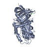
 Movie
Movie Controller

[English] 日本語
 Yorodumi
- PDB-6zrv: Crystal Structure of Stabilized Active Plasminogen Activator Inhi... -
+ Open data

- Basic information
| Entry | Database: PDB / ID: 6zrv | |||||||||
|---|---|---|---|---|---|---|---|---|---|---|
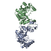
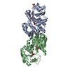
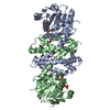
| Title | Crystal Structure of Stabilized Active Plasminogen Activator Inhibitor-1 (PAI-1-W175F) in Complex with an Inhibitory Nanobody (VHH-s-a93, Nb93) | |||||||||
 Components Components |
| |||||||||
 Keywords Keywords | HYDROLASE / plasminogen activator inhibitor-1 / PAI-1 / PAI-1-W175F / serpin / serine protease inhibitor / nanobody / antibody fragment / protein complex | |||||||||
| Function / homology |  Function and homology information Function and homology informationpositive regulation of leukotriene production involved in inflammatory response / negative regulation of smooth muscle cell-matrix adhesion / negative regulation of integrin-mediated signaling pathway / peptidase inhibitor complex / dentinogenesis / positive regulation of coagulation / negative regulation of vascular wound healing / negative regulation of smooth muscle cell migration / Regulation of MITF-M-dependent genes involved in extracellular matrix, focal adhesion and epithelial-to-mesenchymal transition / negative regulation of wound healing ...positive regulation of leukotriene production involved in inflammatory response / negative regulation of smooth muscle cell-matrix adhesion / negative regulation of integrin-mediated signaling pathway / peptidase inhibitor complex / dentinogenesis / positive regulation of coagulation / negative regulation of vascular wound healing / negative regulation of smooth muscle cell migration / Regulation of MITF-M-dependent genes involved in extracellular matrix, focal adhesion and epithelial-to-mesenchymal transition / negative regulation of wound healing / positive regulation of odontoblast differentiation / negative regulation of plasminogen activation / Dissolution of Fibrin Clot / negative regulation of cell adhesion mediated by integrin / positive regulation of monocyte chemotaxis / endopeptidase inhibitor activity / negative regulation of thrombin-activated receptor signaling pathway / negative regulation of blood coagulation / negative regulation of fibrinolysis / positive regulation of blood coagulation / replicative senescence / ECM proteoglycans / negative regulation of endothelial cell apoptotic process / negative regulation of extrinsic apoptotic signaling pathway via death domain receptors / serine protease inhibitor complex / fibrinolysis / negative regulation of proteolysis / BMAL1:CLOCK,NPAS2 activates circadian expression / platelet alpha granule lumen / negative regulation of cell migration / positive regulation of interleukin-8 production / serine-type endopeptidase inhibitor activity / SMAD2/SMAD3:SMAD4 heterotrimer regulates transcription / positive regulation of receptor-mediated endocytosis / positive regulation of angiogenesis / positive regulation of inflammatory response / Platelet degranulation / extracellular matrix / cellular response to lipopolysaccharide / protease binding / angiogenesis / defense response to Gram-negative bacterium / signaling receptor binding / : / extracellular exosome / extracellular region / plasma membrane Similarity search - Function | |||||||||
| Biological species |  Homo sapiens (human) Homo sapiens (human) | |||||||||
| Method |  X-RAY DIFFRACTION / SYNCHROTRON / MOLECULAR REPLACEMENT / Resolution: 1.88 Å X-RAY DIFFRACTION / SYNCHROTRON / MOLECULAR REPLACEMENT / Resolution: 1.88 Å | |||||||||
 Authors Authors | Sillen, M. / Weeks, S.D. / Strelkov, S.V. / Declerck, P.J. | |||||||||
| Funding support |  Belgium, 2items Belgium, 2items
| |||||||||
 Citation Citation | Journal: Int J Mol Sci / Year: 2020 Title: Structural Insights into the Mechanism of a Nanobody That Stabilizes PAI-1 and Modulates Its Activity. Authors: Sillen, M. / Weeks, S.D. / Strelkov, S.V. / Declerck, P.J. | |||||||||
| History |
|
- Structure visualization
| Structure viewer | Molecule: MolmilJmol/JSmol |
|---|
- Downloads & links
-Download
| PDBx/mmCIF format | 6zrv.cif.gz | 115.4 KB | Display | PDBx/mmCIF format |
|---|---|---|---|---|
| PDB format | pdb6zrv.ent.gz | 86.5 KB | Display | PDB format |
| PDBx/mmJSON format | 6zrv.json.gz | Tree view | PDBx/mmJSON format | |
| Others |  Other downloads Other downloads |
-Validation report
| Arichive directory | https://data.pdbj.org/pub/pdb/validation_reports/zr/6zrvftp://data.pdbj.org/pub/pdb/validation_reports/zr/6zrv | HTTPS FTP |
|---|
-Related structure data


-Links
 PDBj
PDBj


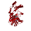
- Assembly
| Deposited unit | 
| ||||||||
|---|---|---|---|---|---|---|---|---|---|
| 1 |
| ||||||||
| Unit cell |
|
-Components
| #1: Protein | Mass: 42780.031 Da / Num. of mol.: 1 / Mutation: W175F Source method: isolated from a genetically manipulated source Source: (gene. exp.) Homo sapiens (human) / Gene: SERPINE1, PAI1, PLANH1 / Plasmid: pETHSUK2 / Production host:  |
|---|---|
| #2: Antibody | Mass: 13252.426 Da / Num. of mol.: 1 Source method: isolated from a genetically manipulated source Source: (gene. exp.) |
| #3: Water | ChemComp-HOH /  Mass: 18.015 Da / Num. of mol.: 315 / Source method: isolated from a natural source / Formula: H2O Mass: 18.015 Da / Num. of mol.: 315 / Source method: isolated from a natural source / Formula: H2O |
| Has protein modification | Y |
-Experimental details
-Experiment
| Experiment | Method: X-RAY DIFFRACTION / Number of used crystals: 1 |
|---|
- Sample preparation
| Crystal | Density Matthews: 2.99 Å3/Da / Density % sol: 58.84 % |
|---|---|
| Crystal grow | Temperature: 293 K / Method: vapor diffusion, sitting drop / pH: 5.5 Details: 1 M DIAMMONIUM PHOSPHATE, O.1 M TRISODIUM CITRATE, 0.2 M SODIUM CHLORIDE |
-Data collection
| Diffraction | Mean temperature: 100 K / Serial crystal experiment: N |
|---|---|
| Diffraction source | Source: SYNCHROTRON / Site: ESRF  / Beamline: ID23-1 / Wavelength: 0.978934 Å / Beamline: ID23-1 / Wavelength: 0.978934 Å |
| Detector | Type: DECTRIS PILATUS3 6M / Detector: PIXEL / Date: Nov 2, 2018 |
| Radiation | Monochromator: Si (111) monochromator / Protocol: SINGLE WAVELENGTH / Monochromatic (M) / Laue (L): M / Scattering type: x-ray |
| Radiation wavelength | Wavelength: 0.978934 Å / Relative weight: 1 |
| Reflection | Resolution: 1.78→83.3 Å / Num. obs: 65134 / % possible obs: 100 % / Redundancy: 13 % / CC1/2: 0.998 / Rmerge(I) obs: 0.126 / Rpim(I) all: 0.036 / Rrim(I) all: 0.131 / Net I/σ(I): 10.9 |
| Reflection shell | Resolution: 1.78→1.88 Å / Redundancy: 12.8 % / Rmerge(I) obs: 2.042 / Num. unique obs: 9357 / CC1/2: 0.697 / Rpim(I) all: 0.589 / Rrim(I) all: 2.127 / % possible all: 100 |
- Processing
| Software |
| ||||||||||||||||||||||||||||||||||||||||||||||||||||||||||||||||||||||||||||||||||||||||||||||||||||||||||||
|---|---|---|---|---|---|---|---|---|---|---|---|---|---|---|---|---|---|---|---|---|---|---|---|---|---|---|---|---|---|---|---|---|---|---|---|---|---|---|---|---|---|---|---|---|---|---|---|---|---|---|---|---|---|---|---|---|---|---|---|---|---|---|---|---|---|---|---|---|---|---|---|---|---|---|---|---|---|---|---|---|---|---|---|---|---|---|---|---|---|---|---|---|---|---|---|---|---|---|---|---|---|---|---|---|---|---|---|---|---|
| Refinement | Method to determine structure: MOLECULAR REPLACEMENT Starting model: 3Q02, 5TP3 Resolution: 1.88→52.69 Å / SU ML: 0.19 / Cross valid method: THROUGHOUT / σ(F): 1.35 / Phase error: 24.81 / Stereochemistry target values: ML
| ||||||||||||||||||||||||||||||||||||||||||||||||||||||||||||||||||||||||||||||||||||||||||||||||||||||||||||
| Solvent computation | Shrinkage radii: 0.9 Å / VDW probe radii: 1.11 Å / Solvent model: FLAT BULK SOLVENT MODEL | ||||||||||||||||||||||||||||||||||||||||||||||||||||||||||||||||||||||||||||||||||||||||||||||||||||||||||||
| Displacement parameters | Biso max: 83.08 Å2 / Biso mean: 36.6323 Å2 / Biso min: 19.24 Å2 | ||||||||||||||||||||||||||||||||||||||||||||||||||||||||||||||||||||||||||||||||||||||||||||||||||||||||||||
| Refinement step | Cycle: final / Resolution: 1.88→52.69 Å
| ||||||||||||||||||||||||||||||||||||||||||||||||||||||||||||||||||||||||||||||||||||||||||||||||||||||||||||
| LS refinement shell | Refine-ID: X-RAY DIFFRACTION / Rfactor Rfree error: 0 / Total num. of bins used: 17 / % reflection obs: 100 %
|
