 Movie
Movie Controller

[English] 日本語
 Yorodumi
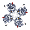
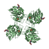
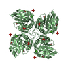
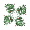
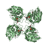
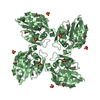
- PDB-6zr0: Crystal structure of tetrameric fibrinogen-like recognition domai... -
+ Open data

- Basic information
| Entry | Database: PDB / ID: 6zr0 | ||||||
|---|---|---|---|---|---|---|---|
| Title | Crystal structure of tetrameric fibrinogen-like recognition domain of FIBCD1 with N-acetylalanine ligand bound | ||||||
 Components Components | Fibrinogen C domain-containing protein 1 | ||||||
 Keywords Keywords | SUGAR BINDING PROTEIN / fibrinogen-like domain / N-acetyl-binding protein | ||||||
| Function / homology |  Function and homology information Function and homology information | ||||||
| Biological species |  Homo sapiens (human) Homo sapiens (human) | ||||||
| Method |  X-RAY DIFFRACTION / SYNCHROTRON / MOLECULAR REPLACEMENT / Resolution: 1.94 Å X-RAY DIFFRACTION / SYNCHROTRON / MOLECULAR REPLACEMENT / Resolution: 1.94 Å | ||||||
 Authors Authors | Shrive, A.K. / Greenhough, T.J. / Williams, H.M. | ||||||
| Funding support |  United Kingdom, 1items United Kingdom, 1items
| ||||||
 Citation Citation | Journal: J.Biol.Chem. / Year: 2023 Title: Crystal structures of human immune protein FIBCD1 suggest an extended binding site compatible with recognition of pathogen associated carbohydrate motifs Authors: Williams, H.M. / Moeller, J.B. / Burns, I. / Schlosser, A. / Sorensen, G.L. / Greenhough, T.J. / Holmskov, U. / Shrive, A.K. #1: Journal: J Biol Chem / Year: 2014Title: Crystal structure of the tetrameric fibrinogen-like recognition domain of fibrinogen C domain containing 1 (FIBCD1) protein Authors: Shrive, A.K. / Moeller, J.B. / Burns, I. / Paterson, J.M. / Shaw, A.J. / Schlosser, A. / Sorensen, G.L. / Greenhough, T.J. / Holmskov, U. | ||||||
| History |
|
- Structure visualization
| Structure viewer | Molecule: MolmilJmol/JSmol |
|---|
- Downloads & links
-Download
| PDBx/mmCIF format | 6zr0.cif.gz | 109.8 KB | Display | PDBx/mmCIF format |
|---|---|---|---|---|
| PDB format | pdb6zr0.ent.gz | 81.7 KB | Display | PDB format |
| PDBx/mmJSON format | 6zr0.json.gz | Tree view | PDBx/mmJSON format | |
| Others |  Other downloads Other downloads |
-Validation report
| Arichive directory | https://data.pdbj.org/pub/pdb/validation_reports/zr/6zr0ftp://data.pdbj.org/pub/pdb/validation_reports/zr/6zr0 | HTTPS FTP |
|---|
-Related structure data
| Related structure data | 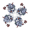 6zqrC 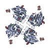 6zqxC 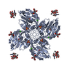 6zqyC  6zr3C 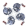 6zr4C  4m7hS C: citing same article ( S: Starting model for refinement |
|---|---|
| Similar structure data | |
| Experimental dataset #1 | Data reference: 10.21252/zcfy-cw20 / Data set type: diffraction image data / Details: https://doi.org/10.21252/zcfy-cw20 |


-Links
 PDBj
PDBj


- Assembly
| Deposited unit | 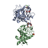
| ||||||||
|---|---|---|---|---|---|---|---|---|---|
| 1 | 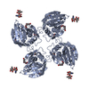
| ||||||||
| 2 | 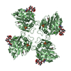
| ||||||||
| Unit cell |
|
-Components
-Protein / Sugars , 2 types, 3 molecules AB
| #1: Protein | Mass: 25688.055 Da / Num. of mol.: 2 / Fragment: fibrinogen-like recognition domain Source method: isolated from a genetically manipulated source Source: (gene. exp.) Homo sapiens (human) / Gene: FIBCD1, UNQ701/PRO1346 / Plasmid: pNT-Bac / Production host:   Spodoptera frugiperda (fall armyworm) / References: UniProt: Q8N539 Spodoptera frugiperda (fall armyworm) / References: UniProt: Q8N539#2: Polysaccharide | alpha-L-fucopyranose-(1-3)-2-acetamido-2-deoxy-beta-D-glucopyranose | |
|---|
-Non-polymers , 6 types, 259 molecules 


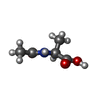

| #3: Chemical |  Mass: 40.078 Da / Num. of mol.: 2 / Source method: obtained synthetically / Formula: Ca Mass: 40.078 Da / Num. of mol.: 2 / Source method: obtained synthetically / Formula: Ca#4: Chemical |  Mass: 96.063 Da / Num. of mol.: 3 / Source method: obtained synthetically / Formula: SO4 Mass: 96.063 Da / Num. of mol.: 3 / Source method: obtained synthetically / Formula: SO4#5: Chemical | ChemComp-GOL / |  Mass: 92.094 Da / Num. of mol.: 1 Mass: 92.094 Da / Num. of mol.: 1Source method: isolated from a genetically manipulated source Formula: C3H8O3 #6: Chemical |  Mass: 60.052 Da / Num. of mol.: 3 Mass: 60.052 Da / Num. of mol.: 3Source method: isolated from a genetically manipulated source Formula: C2H4O2 #7: Chemical |  Type: L-peptide linking / Mass: 131.130 Da / Num. of mol.: 2 Type: L-peptide linking / Mass: 131.130 Da / Num. of mol.: 2Source method: isolated from a genetically manipulated source Formula: C5H9NO3 / Feature type: SUBJECT OF INVESTIGATION #8: Water | ChemComp-HOH / | Mass: 18.015 Da / Num. of mol.: 248 / Source method: isolated from a natural source / Formula: H2O |
|---|
-Details
| Has ligand of interest | Y |
|---|---|
| Has protein modification | Y |
-Experimental details
-Experiment
| Experiment | Method: X-RAY DIFFRACTION / Number of used crystals: 1 |
|---|
- Sample preparation
| Crystal | Density Matthews: 3.06 Å3/Da / Density % sol: 59.75 % |
|---|---|
| Crystal grow | Temperature: 293 K / Method: vapor diffusion, sitting drop / pH: 6.5 Details: 1.5 M Ammonium Sulphate, 8% Dioxane, 0.1 M MES pH 6.5 |
-Data collection
| Diffraction | Mean temperature: 100 K / Serial crystal experiment: N |
|---|---|
| Diffraction source | Source: SYNCHROTRON / Site: Diamond / Beamline: I04 / Wavelength: 0.9795 Å |
| Detector | Type: DECTRIS PILATUS 6M-F / Detector: PIXEL / Date: Jun 30, 2016 |
| Radiation | Protocol: SINGLE WAVELENGTH / Monochromatic (M) / Laue (L): M / Scattering type: x-ray |
| Radiation wavelength | Wavelength: 0.9795 Å / Relative weight: 1 |
| Reflection | Resolution: 1.94→84.44 Å / Num. obs: 46069 / % possible obs: 99.1 % / Redundancy: 3.2 % / Rmerge(I) obs: 0.08 / Net I/σ(I): 9.4 |
| Reflection shell | Resolution: 1.94→1.99 Å / Redundancy: 3.2 % / Rmerge(I) obs: 0.401 / Num. unique obs: 3105 / % possible all: 98.8 |
- Processing
| Software |
| |||||||||||||||||||||||||||||||||||||||||||||||||||||||||||||||||||||||||||||||||||||||||||||||||||||||||||||||||||||||||||||||||||||||||||||||||
|---|---|---|---|---|---|---|---|---|---|---|---|---|---|---|---|---|---|---|---|---|---|---|---|---|---|---|---|---|---|---|---|---|---|---|---|---|---|---|---|---|---|---|---|---|---|---|---|---|---|---|---|---|---|---|---|---|---|---|---|---|---|---|---|---|---|---|---|---|---|---|---|---|---|---|---|---|---|---|---|---|---|---|---|---|---|---|---|---|---|---|---|---|---|---|---|---|---|---|---|---|---|---|---|---|---|---|---|---|---|---|---|---|---|---|---|---|---|---|---|---|---|---|---|---|---|---|---|---|---|---|---|---|---|---|---|---|---|---|---|---|---|---|---|---|---|---|
| Refinement | Method to determine structure: MOLECULAR REPLACEMENT Starting model: PDBID 4M7H Resolution: 1.94→84.44 Å / Cor.coef. Fo:Fc: 0.949 / Cor.coef. Fo:Fc free: 0.943 / SU B: 3.109 / SU ML: 0.088 / Cross valid method: THROUGHOUT / σ(F): 0 / ESU R: 0.138 / ESU R Free: 0.123 / Stereochemistry target values: MAXIMUM LIKELIHOOD Details: HYDROGENS HAVE BEEN ADDED IN THE RIDING POSITIONS U VALUES : REFINED INDIVIDUALLY
| |||||||||||||||||||||||||||||||||||||||||||||||||||||||||||||||||||||||||||||||||||||||||||||||||||||||||||||||||||||||||||||||||||||||||||||||||
| Solvent computation | Ion probe radii: 0.8 Å / Shrinkage radii: 0.8 Å / VDW probe radii: 1.2 Å / Solvent model: MASK | |||||||||||||||||||||||||||||||||||||||||||||||||||||||||||||||||||||||||||||||||||||||||||||||||||||||||||||||||||||||||||||||||||||||||||||||||
| Displacement parameters | Biso max: 62.83 Å2 / Biso mean: 21.69 Å2 / Biso min: 12.45 Å2
| |||||||||||||||||||||||||||||||||||||||||||||||||||||||||||||||||||||||||||||||||||||||||||||||||||||||||||||||||||||||||||||||||||||||||||||||||
| Refinement step | Cycle: final / Resolution: 1.94→84.44 Å
| |||||||||||||||||||||||||||||||||||||||||||||||||||||||||||||||||||||||||||||||||||||||||||||||||||||||||||||||||||||||||||||||||||||||||||||||||
| Refine LS restraints |
| |||||||||||||||||||||||||||||||||||||||||||||||||||||||||||||||||||||||||||||||||||||||||||||||||||||||||||||||||||||||||||||||||||||||||||||||||
| LS refinement shell | Resolution: 1.94→1.99 Å / Rfactor Rfree error: 0 / Total num. of bins used: 20
|
